آخر المواضيع المضافة

 النبات
الحيوان
الأحياء المجهرية
علم الأمراض
التقانة الإحيائية
التقنية الحيوية المكروبية
التقنية الحياتية النانوية
علم الأجنة
الأحياء الجزيئي
علم وظائف الأعضاء
الغدد
المضادات الحيوية
النبات
الحيوان
الأحياء المجهرية
علم الأمراض
التقانة الإحيائية
التقنية الحيوية المكروبية
التقنية الحياتية النانوية
علم الأجنة
الأحياء الجزيئي
علم وظائف الأعضاء
الغدد
المضادات الحيوية| Viral Genomes Are Packaged into Their Coats |


|
|
Read More
Date: 22-12-2015
Date: 6-12-2015
Date: 27-11-2015
|
Viral Genomes Are Packaged into Their Coats
KEY CONCEPTS
-The length of DNA that can be incorporated into a virus is limited by the structure of the head shell.
-Nucleic acid within the head shell is extremely condensed.
-Filamentous RNA viruses condense the RNA genome as they assemble the head shell around it.
-Spherical DNA viruses insert the DNA into a preassembled protein shell.
From the perspective of packaging the individual sequence, there is an important difference between a cellular genome and a virus. The cellular genome is essentially indefinite in size; the number and location of individual sequences can be changed by duplication, deletion, and rearrangement. Thus, it requires a generalized method for packaging its DNA—one that is insensitive to the total content or distribution of sequences. By contrast, two restrictions define the needs of a virus. The amount of nucleic acid to be packaged is predetermined by the size of the genome, and it must all fit within a coat assembled from a protein or proteins coded by the viral genes.
A virus particle is deceptively simple in its superficial appearance. The nucleic acid genome is contained within a capsid, which is a symmetrical or quasisymmetrical structure assembled from one or only a few proteins. Attached to the capsid (or incorporated into it) are other structures; these structures are assembled from distinct proteins and are necessary for infection of the host cell.
The virus particle is tightly constructed. The internal volume of the capsid is rarely much greater than the volume of the nucleic acid it must hold. The difference is usually less than twofold, and often the internal volume is barely larger than the nucleic acid.
In its most extreme form, the restriction that the capsid must be assembled from proteins encoded by the virus means that the entire shell is constructed from a single type of subunit. The rules for assembly of identical subunits into closed structures restrict the capsid to one of two types. For the first type, the protein subunits stack sequentially in a helical array to form a filamentous or rodlike shape. For the second type, they form a pseudospherical shell—a type of structure that conforms to a polyhedron with icosahedral symmetry. Some viral capsids are assembled from more than a single type of protein subunit. Although this extends the exact types of structures that can be formed, most viral capsids conform to the general classes of quasicrystalline filaments or icosahedrons.
There are two general solutions to the problem of how to construct a capsid that contains nucleic acid:
- The protein shell can be assembled around the nucleic acid, thereby condensing the DNA or RNA by protein–nucleic acid interactions during the process of assembly.
-The capsid can be constructed from its component(s) in the form of an empty shell, into which the nucleic acid must be inserted, being condensed as it enters.
The capsid is assembled around the genome for single-stranded RNA viruses. The principle of assembly is that the position of the RNA within the capsid is determined directly by its binding to the proteins of the shell. The best characterized example is tobacco mosaic virus (TMV). Assembly begins at a duplex hairpin that lies within the RNA sequence. From this nucleation center, assembly proceeds bidirectionally along the RNA until it reaches the ends.
The unit of the capsid is a two-layer disk, with each layer containing 17 identical protein subunits. The disk is a circular structure, which forms a helix as it interacts with the RNA. At the nucleation center, the RNA hairpin inserts into the central hole in the disk, and the disk changes conformation into a helical structure that surrounds the RNA. Additional disks are added, with each new disk pulling a new stretch of RNA into its central hole. The RNA becomes coiled in a helical array on the inside of the protein shell, as illustrated in FIGURE .1.

FIGURE .1 A helical path for TMV RNA is created by the stacking of protein subunits in the virion (the entire virus particle).
The spherical capsids of DNA viruses are assembled in a different way, as best characterized for the phages lambda and T4. In each case, an empty head shell is assembled from a small set of proteins. The duplex genome then is inserted into the head, accompanied by a structural change in the capsid.
FIGURE .2 summarizes the assembly of lambda. It begins with a small head shell that contains a protein “core.” This is converted to an empty head shell of more distinct shape. At this point the DNA packaging begins, the head shell expands in size (though it remains the same shape), and finally the full head is sealed by the addition of the tail.
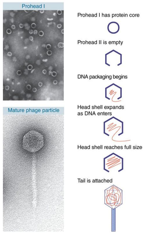
FIGURE .2 Maturation of phage lambda passes through several stages. The empty head changes shape and expands when it becomes filled with DNA, diagrammed on the left. The electron micrographs on the right show the particles at the beginning (top) and the end (bottom) of the maturation pathway.
Top photo reproduced from: Cue, D., and Feiss M. 1993. Proc Natl Acad Sci USA 90: 9240–9294. Copyright © 2004 National Academy of Sciences, U.S.A. Bottom photo courtesy of Robert Duda, University of Pittsburgh.
A double-stranded DNA that spans short distances is a fairly rigid rod, yet it must be compressed into a compact structure to fit within the capsid. This packaging can be achieved by a smooth coiling of the DNA into the head or it might require introduction of abrupt bends.
Inserting DNA into a phage head involves two types of reaction: translocation and condensation. Both are energetically unfavorable.
Translocation is an active process in which the DNA is driven into the head by an ATP-dependent mechanism. A common mechanism for translocation is used for many viruses that replicate by a rolling circle mechanism to generate long tails that contain multimers of the viral genome. The best characterized example is phage lambda. The genome is packaged into the empty capsid by the terminase enzyme. FIGURE .3 summarizes the process.
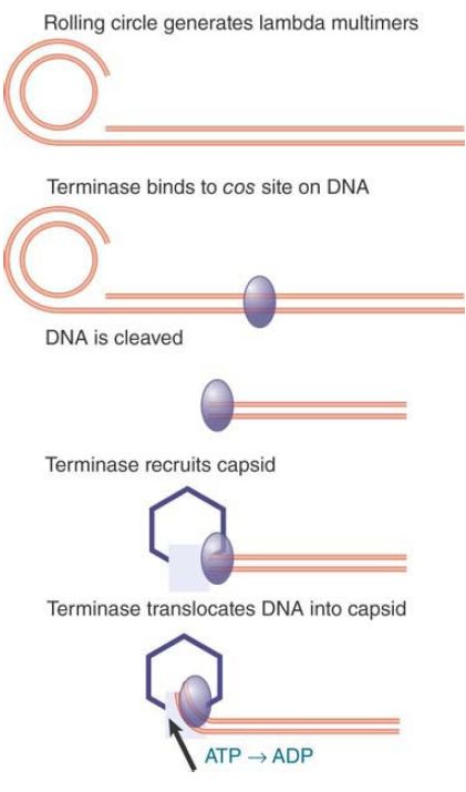
FIGURE .3 Terminase protein binds to specific sites on a multimer of virus genomes generated by rolling circle replication. It cuts the DNA and binds to an empty virus capsid, and then uses energy from hydrolysis of ATP to insert the DNA into the capsid.
The terminase was first recognized (and named) for its role in generating the ends of the linear phage DNA by cleaving at cos sites. (The name cos reflects the fact that it generates cohesive ends that have complementary single-stranded tails.) The phage genome encodes two subunits that make up the terminase. One subunit binds to a cos site; at this point it is joined by the other subunit, which cuts the DNA. The terminase assembles into a heterooligomer in a complex that also includes integration host factor (IHF; a dimer that is encoded by the bacterial genome). It then binds to an empty capsid and uses ATP hydrolysis to power translocation along the DNA. The translocation drives the DNA into the empty capsid.
Another method of packaging uses a structural component of the phage. In the Bacillus subtilis phage ϕ29, the motor that inserts the DNA into the phage head is an integral structure that connects the head to the tail. It functions as a rotary motor, where the motor action effects the linear translocation of the DNA into the phage head. The same motor is used to eject the DNA from the phage head when it infects a bacterium.
Less is known about the mechanism(s) of condensation into an empty capsid, except that capsids typically contain “internal proteins” as well as DNA. Such internal proteins might provide some sort of scaffolding onto which the DNA condenses. This would be similar to the use of the proteins of the shell in the plant RNA viruses (e.g., TMV, described earlier in this section).
How specific is the packaging? It cannot depend simply on particular sequences, because deletions, insertions, and substitutions all fail to interfere with the assembly process. The relationship between DNA and the head shell has been investigated directly by determining which regions of the DNA can be chemically crosslinked to the proteins of the capsid. The surprising answer is that all regions of the DNA are more or less equally susceptible.
This probably means that when DNA is inserted into the head it follows a general rule for condensing, but the pattern is not determined by particular sequences.
These varying mechanisms of virus assembly all accomplish the same end: packaging a single DNA or RNA molecule into the capsid. Some viruses, however, have genomes that consist of multiple nucleic acid molecules. Reovirus contains 10 doublestranded RNA segments, all of which must be packaged into the capsid. Specific sorting sequences in the segments might be required to ensure that the assembly process selects one copy of each different molecule in order to collect a complete set of genetic information. In the simpler case of phage ϕ6, which packages three different segments of double-stranded RNA into one capsid, the RNA segments must bind in a specific order; as each is incorporated into the capsid, it triggers a change in the
conformation of the capsid that creates binding sites for the next segment.
Some plant viruses are multipartite: Their genomes consist of segments, each of which is packaged into a different capsid. An example is alfalfa mosaic virus (AMV), which has four different single-stranded RNAs, each of which is packaged independently into a coat comprising the same protein subunit. A successful infection depends on the entry of one of each type into the cell. The four components of AMV exist as particles of different sizes. This means that the same capsid protein can package each RNA into its own characteristic particle. This is a departure from the packaging of a unique length of nucleic acid into a capsid of fixed shape.
The assembly pathway of viruses whose capsids have only one authentic form might be diverted by mutations that cause the formation of aberrant monster particles in which the head is longer than usual. These mutations show that a capsid protein(s) has an intrinsic ability to assemble into a particular type of structure, but the exact size and shape can vary.
Some of the mutations occur in genes that code for assembly factors, which are needed for head formation, but are not themselves part of the head shell. Such ancillary proteins limit the
options of the capsid protein, reducing variation in the assembly pathway. Comparable proteins are employed in the assembly of cellular chromatin .


|
|
|
|
الصين.. طريقة لمنع تطور قصر النظر لدى تلاميذ المدارس
|
|
|
|
|
|
|
ماذا سيحدث خلال كسوف الشمس يوم السبت؟
|
|
|
|
|
|
|
قسم الشؤون الدينية يختتم محاضراته الرمضانية في صحن مرقد أبي الفضل العباس (عليه السلام)
|
|
|