آخر المواضيع المضافة

 النبات
الحيوان
الأحياء المجهرية
علم الأمراض
التقانة الإحيائية
التقنية الحيوية المكروبية
التقنية الحياتية النانوية
علم الأجنة
الأحياء الجزيئي
علم وظائف الأعضاء
الغدد
المضادات الحيوية
النبات
الحيوان
الأحياء المجهرية
علم الأمراض
التقانة الإحيائية
التقنية الحيوية المكروبية
التقنية الحياتية النانوية
علم الأجنة
الأحياء الجزيئي
علم وظائف الأعضاء
الغدد
المضادات الحيوية| Contact Inhibition |


|
|
Read More
Date: 19-12-2015
Date: 17-11-2020
Date: 10-5-2016
|
Contact Inhibition
Cultured cells, particularly when grown as a monolayer attached to a substrate, will go through a reproducible growth cycle following subculture (1) (see Cell Line). The phases normally defined are (1) the lag phase, before the culture starts to proliferate; (2) the exponential , or log , phase, when the cells are dividing rapidly and the population doubles over a fixed time; and (3) the plateau phase, when the cell number remains stable with further time in culture. As a culture reaches the end of the exponential phase and enters plateau, it responds to cell contact, reduced substrate availability, a reduction in nutrients, and a number of other stimuli that inhibit a further increase in cell numbers. These have been divided into two major components: contact inhibition and density limitation of cell proliferation . Contact inhibition is the cessation of cell motility that occurs when a cell culture reaches confluence. It was first described from observations of randomly migrating fibroblasts made in time-lapse cinemicrography (2). Normal fibroblasts cultured at low cell density show a spindle-shaped morphology, with a reversible polarity along their long axis, which is also the direction of cell migration. When the cell encounters another similar cell, contact induces a reversal of the polarity; the ruffling in the leading tip of the cell ceases and transfers to the opposite tip, which then becomes the anterior end, and migration resumes in the opposite direction. When contact is made with another cell, the process of reversal of polarity is repeated, unless the cell is surrounded by other cells, when membrane ruffling ceases and cell migration is inhibited. In the cases of normal human or chick diploid fibroblasts, the cell assumes a parallel array that gives the appearance of whorls in the cell monolayer under low power microscopy or naked eye observation
At the time that motility ceases, the cells are still capable of proliferation, but the culture becomes crowded following one or two population doublings, the cells assume a narrower spindle shape, and cell spreading is reduced beyond the point where the cell is able to re-enter the cell cycle (3). At this point, proliferation ceases in a normal cell culture, and the culture remains as a monolayer. Further growth is inhibited by density limitation of cell proliferation , and the culture enters the plateau phase of the growth cycle. Density limitation of cell proliferation is distinct from contact inhibition, which affects primarily membrane ruffling, polarity, and cell migration. The mechanisms are, however, similar, although the detail remains obscure. Subconfluent cells have many more adhesions to the substrate than to other cells, favoring motility and proliferation. When the culture becomes confluent, the number of cell–cell adhesions (cadherins) increases, but they are still exceeded by cell–substrate adhesions (integrins), which inhibit cell motility but not proliferation. When the culture becomes crowded, the number of cell–cell adhesions increases, and cell–substrate adhesions are reduced, maintaining inhibition of motility but now inhibiting re-entry into the cell cycle. As contact with adjacent cells and contact with the substrate are mediated via different classes of receptors (4), the signaling that results from activation of these receptors is potentially different, although exactly how remains unclear.
In addition to alterations in cell spreading, adhesion receptors, and the actin cytoskeleton when cells reach a high density, the cells are also exposed to reduced nutrient levels, due to a higher rate of utilization, and increased levels of catabolites, both of which will tend to inhibit proliferation. In a static culture, this may generate a depleted layer above the cells, across which nutrients and growth factors must diffuse to reach the cells. The diffusion rate will be significantly slower with higher molecular weight components of the medium, including growth factors and peptide hormones such as insulin. Support for this diffusion boundary layer hypothesis was provided by experiments where local irrigation was increased, by means of a micropump, and resulted in a localized increase in the frequency of labeling with [3H]-thymidine (5). As the range of the diffusion boundary was short, it was proposed that the limitation was principally in growth factors, depleted by binding to cell-surface receptors and endocytosis, which was confirmed by the observation that addition of growth factors or serum to the medium will induce further proliferation (6).
On the other hand, one of the main observations that confirms that contact inhibition and density limitation of cell proliferation are not simply evidence of environmental deterioration is the result of the so-called “wounded monolayer” experiment (7). If a confluent, growth-arrested monolayer is scored with a sharp instrument, creating a bare patch devoid of cells, the surrounding cells respond as follows. The edge cells start to show ruffling of the cell membrane bordering the wound, spread out, and migrate into the space. When they have spread beyond a critical point, they enter the cell cycle and divide, and they continue to do so until the space of the wound is filled. They then stop migrating, but continue to proliferate until the cell density in the wound matches the crowded state of the rest of the monolayer; at this point they stop dividing. Meanwhile, no other cells distant from the wound show any sign of entering the cell cycle. This confirms that a major component of density limitation of cell proliferation is due to geometry and cell shape, rather than simply nutritional or growth factor limitation.
If a culture of normal fibroblasts that is growth arrested at high density is fed with medium containing serum or growth factors, many of the cells will reenter the cell cycle, resulting in the formation of a second cell layer over the first; if this is repeated, multilayered cultures can be produced. It has been proposed that the first layer of cells secretes a collagen overlay (which may produce a further diffusion barrier) and the second cell layer grows on top of this, technically not disobeying the rules of contact inhibition and density limitation of cell proliferation. This second layer, however, is still limited by contact and density, and when it reaches confluence, it will form a second layer of parallel arrays of cells, not colinear with the first. If perfused, these cultures may continue to increase up to 20 or 30 cell layers deep (8).
When cultures of normal epithelium reach confluence, they also become contact-inhibited. In less dense cultures, however, they do not show the random migration seen in cultures of fibroblasts, but tend to grow in patches with ruffling of the membrane, cell spreading, and proliferation restricted to the outer edge of the patch (9). Their motility is limited, but when it occurs, it tends to involve the whole patch, which moves as a unit. Multilayering in normal epithelial cultures also occurs, but tends to imply maturation perpendicular to the substrate, rather than overgrowth. Epidermal cells, for example, will become stratified, with the upper layers of cells becoming progressively more keratinized. This is accentuated if the cells are grown on collagen, particularly in the presence of dermal fibroblasts and due to integrin down-regulation. Epithelial cells, which are derived from simple epithelium that is only one cell thick in vivo, tend to be obligate monolayers in culture. Likewise, normal endothelium from the lining of blood vessels will not pile up in culture, although after some time at high density it will tend to curl up and form secondary structures resembling capillaries (10), particularly if grown on an extracellular matrix, such as Matrigel.
Transformation allows cells to escape from the restrictions of contact inhibition and density limitation of cell proliferation. On one hand, they often lack, or have modified, the appropriate receptors or cell-adhesion molecules to recognize each other on contact; on the other hand, they are no longer dependent on cell spreading to allow entry into the cell cycle. Many transformed cell lines will propagate in suspension, without any substrate attachment. In addition, transformed cells often produce autocrine growth factors or have permanently active steps in the signal transduction cascades that promote cell proliferation (11-13). Hence, transformed cells will grow to a higher density and form multilayered cultures quite readily. The limitations imposed by density now tend to be diffusion-related, principally nutrient, catabolite, and gaseous (dissolved O2 in and CO2 out). One major limitation is the release of lactic acid by the cells, as they are generally more anaerobic in their metabolism than normal cells, and this depresses the pH, initially inhibiting proliferation and ultimately killing the cells. Although transformed cell cultures enter a plateau phase of culture when they reach a high density, the growth fraction in transformed cultures in plateau can be quite high (10–20%), unlike the plateau in normal cell cultures, where the growth fraction is very low (<5%). A steady state is reached when cell proliferation is matched by cell necrosis or apoptosis, and the cells deteriorate rapidly and irreversibly if the medium is not replenished.
The differences in response to high cell density between normal and transformed cells have been exploited in the isolation of transformed foci. If a monolayer of 3T3 cells is transfected with an oncogene, especially in the exponential phase of growth, when transfection is more efficient, it will show foci of transformed cells when the culture reaches confluence, because of the lack of contact inhibition of cell motility and density limitation of cell proliferation in successfully transformed cells (14). Likewise, transformed cells plated on a confluent monolayer will continue to grow and may form foci, or they may infiltrate the monolayer. Normal cells plated on a confluent monolayer will give a varying response, depending on the cell lineage of the confluent monolayer. If the plated normal cells are of the same lineage as the confluent monolayer, they will not grow; for instance, fibroblasts will not grow on a confluent monolayer of fibroblasts, and normal glial cells will not grow on a confluent monolayer of normal glial cells (15), whereas their transformed counterparts will. On the other hand, normal glial cells will grow on a confluent monolayer of fibroblasts, and normal keratinocytes will grow on a monolayer of normal 3T3 cells (16).
Cultures that are propagated in suspension will, of course, not encounter contact inhibition. Nevertheless, they are subject to density limitation of cell proliferation. Part of this is undoubtedlydue to nutrient depletion, catabolite buildup, and pH depression, but even if limiting nutrients are replaced and the pH is stabilized, high density suspension cultures do not increase significantly above 1–2 × 10⁶ cells/ml. Quite often suspension cultures, such as hybridomas, will remain in the plateau phase for only a short time and then deteriorate rapidly, leaving few viable cells in the culture. The reason for this appears to be that the cells tend to enter apoptosis when they reach a high density. Some moderate success has been achieved overexpressing bcl in these cells to inhibit apoptosis (17).
References
1. R. I. Freshney (1994) Culture of Animal Cells, a Manual of Basic Technique, Wiley-Liss, New York, pp. 153–157.
2. M. Abercrombie and J. E. M. Heaysman (1954) Exp. Cell Res. 6, 293–306.
3. I. Folkman and A. Moscona, (1978) Nature 273, 345–349.
4. S. Levenberg, B.-Z. Katz, K. M. Yamada, and B. Geiger (1998) J. Cell Sci. 111 347–357.
5. M. G. P. Stoker (1973) Nature 246, 200–203.
6. G. A. Dunn and G. W. Ireland (1984) Nature 312, 63–65.
7. B. Alberts, D. Bray, J. Lewis, M. Raff, K. Roberts, and J. D. Watson (1989) Molecular Biology of the Cell, 3rd ed., Garland Publishing, New York, p. 898.
8. P. F. Kruse Jr. and E. Miedema (1965) J. Cell Biol. 27, 273.
9. I. McKay and J. Taylor-Papadimitriou (1981) Exp. Cell Res. 134, 465–470.
10. J. Folkman and C. Haudenschild (1980) Nature 288, 551–556.
11. P. Kahn and T. Graf (1986) Oncogenes and Growth Control, Springer, Berlin.
12. K. Siegfried, Y. H. Han, M. A. A. DeMichele, J. D. Hunt, A. L. Gaither, and F. Cuttitta (1994( J. Biol. Chem. 269, 8596–8603.
13. A. Balmain and K. Brown (1988) Adv. Cancer Res. 51, 147–182.
14. R. I. Freshney (1994) Culture of Animal Cells, a Manual of Basic Technique, Wiley-Liss, New York, p. 233.
15. C. M. MacDonald, R. I. Freshney, E. Hart, and D. I. Graham (1985) Exp. Cell Biol. 53, 130–137.
16. J. G. Rheinwald and H. Green (1975) Cell 6, 331–344.
17. S. Terada, Y. Itoh, H. Ueda, and E. Suzuki (1997) Cytotechnology 24, 135–141.


|
|
|
|
صنع الذكريات والتفكير يدمر الدماغ.. دراسة تشرح السبب
|
|
|
|
|
|
|
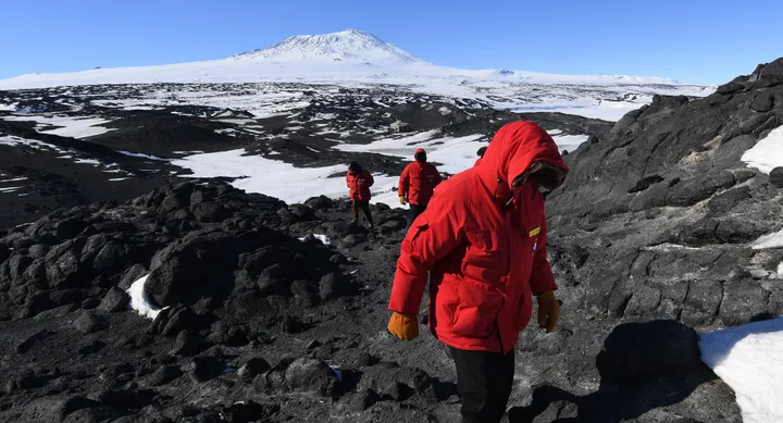
بركان ينفت الذهب في أقصى جنوب الأرض.. ما القصة؟
|
|
|
|
|
|
السيد الصافي يزور قسم التربية والتعليم ويؤكد على دعم العملية التربوية للارتقاء بها
|
|
|
|
لمنتسبي العتبة العباسية قسم التطوير ينظم ورشة عن مهارات الاتصال والتواصل الفعال
|
|
|
|
في جامعة بغداد.. المؤتمر الحسيني الثاني عشر يشهد جلسات بحثية وحوارية
|
|
|
|
للأطفال نصيبهم من جناح جمعية العميد في معرض تونس الدولي للكتاب.. عمّ يبحثون؟
|