


 النبات
 الحيوان
 الأحياء المجهرية
 علم الأمراض
 التقانة الإحيائية
 التقنية الحيوية المكروبية
 التقنية الحياتية النانوية
 علم الأجنة
 الأحياء الجزيئي
 علم وظائف الأعضاء
 الغدد
الغدد
 المضادات الحيوية
|
Read More
Date: 9-7-2021
Date: 12-7-2021
Date: 16-7-2021
|
Skeletal system
The skeletal system is constituted by bones, cartilages and ligaments. This system provides ‘the shape’ to the body. Further, bones remain as regions for the attachment of muscles. It also helps to hold weight. Structures like skull protect inner organs. This system is also useful in locomotion. The bones remain as reservoirs of fat and certain minerals. The bone marrow is the site for the production of erythrocytes.
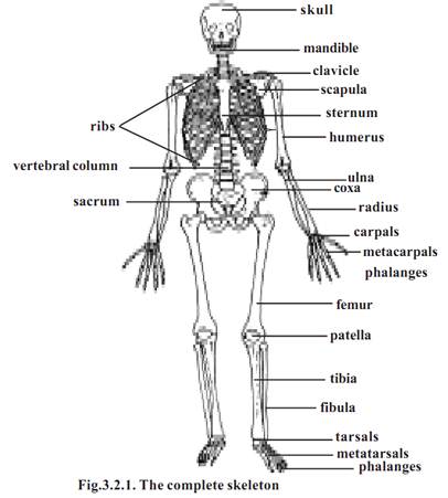
The bones can be long, short, flat or irregular in shape. Hands and legs have long bones. Short bones are broad in shape. Carpals (wrist bones) and tarsals (antkle bones) are shorter. Flat bones are thin and flattened. Skull bones, ribs, sternum and scapula (shoulder blade) are flat bones. Vertebral and facial bones are irregular in shape.
Structure of a typical long bone
A bone is covered by a double layered sheath called the periosteum. The outer layer of the periosteum is fibrous in nature. It is a dense collagenous layer having blood vessels and nerves.
A growing long bone has three regions. The long bony part is the dia- physis or shaft. It is made up of compact bone.
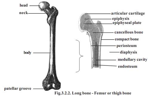
The end of the bone consists of epiphysis. It is made up of spongy bone. The outer surface of epiphysis is formed of compact bone. In between the epiphysis and diaphysis epiphyseal or growth plate is found. It is made up of hyaline cartilage. Growth in length of bone occurs at this plate.
The cavity inside the diaphysis is called the medullary cavity. This cavity is lined by a membrane called the endosteum. The cavity inside the diaphysis in adults contain yellow marrow. It is mostly adipose tissue. The medullary cavity of the epiphysis contains red marrow concerned with blood cell formation.
Dried, prepared bones are used to study skeletal anatomy. The bones are named according to their position in the body. The named bones are divided into two categories: (1) the axial skeleton and (2) the appendicular skeleton. The axial skeleton consists of the skull, hyoid bone, vertebral column and thoracic cage. The appendicular skeleton consists of the limbs and their girdles. In human body, there are 206 bones, of these 80 are in the axial skeleton, 126 in the appendicular skeleton. Among the bones of the axial skeleton 28 bones are in the skull, 26 bones in the vertebral column, 25 bones in the thoracic cage and one remains as the hyoid bone. (Details as found below)
Axial skeleton - It forms the upright axis of the body. It protects the brain, the spinal cord and the vital organs found within the thorax.
a) Skull - The human cranial capacity is about 1500 cm3. It consists of 22 bones. It protects the brain. It supports the organs of vision, hearing, smell and taste. The lower jaw or mandible remains specially attached to the skull. The skull or cranium is covered by eight bones. They are one pair each of parietal and temporal, individual bones as frontal, sphenoid, occipital and ethmoid. These bones are joined by sutures to form a compact box like structure. The sutures are immovable
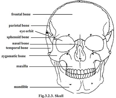
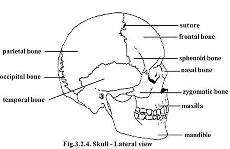
In the front there are 14 facial bones. Of this maxilla, zygomatic, palatine, lacrymal, nasal and inferior nasal koncha remain as pairs. Mandible or lower jaw and vomer are unpaired bones.
The parietal and occipital bones are major bones on the posterior side of the skull. The parietal bones are joined to the occipital bone at the back. The side of the head is formed of the parietal and the temporal bones. The large hole in the temporal bone is the external auditory meatus. This opening is meant for transmitting sound waves towards the eardrum. On the lateral side immediately anterior to the temporal, the sphenoid bone is seen. Anterior to the sphenoid bone is the zygomatic bone or cheek bone. It is a prominent bone on the face. The upper jaw is formed of the maxilla. The mandible constitutes the lower jaw.
The major bones seen from the frontal view are the frontal bone, zygomatic bone the maxillae and the mandible. The most prominent openings in the skull are the orbits and the nasal cavity. The two orbits are meant for accommodating the eyes. The bones of the orbits provide protection for the eyes and attachment points for the muscles that move the eyes. The bones forming the oribits are the frontal, sphenoid, zygomatic, maxilla, lacrymal, ethmoid and palatine. The head region also contains 6 ear ossicles. They are Malleus (2), incus (2) and stapes (2).
A large opening found at the base of the skull is the foramen magnum. Through this opening the medulla oblongata of the brain descends down as the spinal cord.
b) Vertebrae - The vertebrae make up the slight S-shaped vertebral column or backbone. The vertebral column consists of 26 bones. They are divided into 5 regions. They are the cervical (7), thoracic (12), lumbar (5), sacral (1) and coccygeal (1) vertebrae.
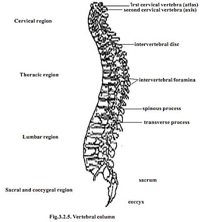
Vertebra - Structure: The main load - bearing portion of a vertebra is a solid disc of bone called the centrum. The central of adjacent vertebrae are separated by intervertebral discs of cartilage. Projecting from the centrum dorsally is a vertebral arch. It encloses the neural canal. This canal contains the spinal cord. Several bony projections are seen on the vertebral arch. On of muscles. Further, there are two superior and two inferior processes meant for articulation with the neighboring vertebra.
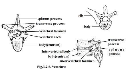
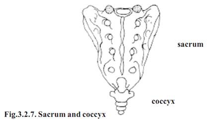
The first cervical vertebra is the atlas. It balances and supports the head. It has no centrum. The second is the axis. The sacral vertebrae are fused. They form a triangular structure called the sacrum. The coccygeal vertelera has no function. It is a vestige. During development in the embryonic stage there are nearby 34 vertebrae present. Of these, 5 sacral bones are fused to form a single sacral bone. 4 or 5 coccygeal bones are fused to form a single coccyx.
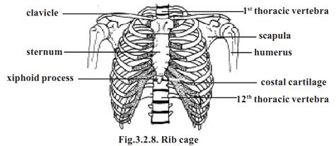
c) Rib cage - There are 12 pairs of ribs. Each articulates with a thoracic vertebra. In the front, the first ten pairs are attached to the sternum (breast bone) by costal cartilages. The first seven are attached directly to the ster-num. They are called the true ribs. Cartilages of 8th, 9th and 10th ribs are fused and attached to 7th. They are called the false ribs. 11th 12th pairs are not attached to the sternum. They are called floating ribs.
Appendicular skeleton
It consists of the bones of the upper and lower limbs and the girdles by which they are attached to the body.
Pectoral girdle - The hands are attached to the pectoral girdle. Both of them are attached loosely by muscles to the body. This arrangement facilitates freedom of movement. Hence it is possible to place the hand in a wide range of positions.
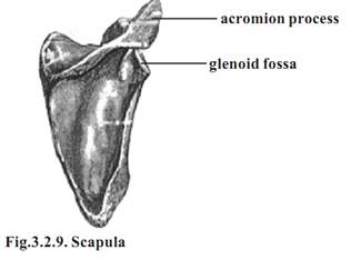
The pectoral or shoulder girdle consists of two pairs of bones. Each pair has a scapula or shoulder blade and a clavicle or collarbone. The scapula is a flat, triangular bone. A glenoid fossa is located in the superior lateral region of the scapula. It articulates with the head of the humerus. The clavicle is a long bone. It has a slight S-shaped curve. It can be easily seen and felt. The clavicle holds the upper limb away from the body.
Pelvic girdle or pelvis - It is a ring of bones formed by the sacrum and paired bones called the coxae or hip bones.
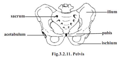
Each coxa is formed by the fusion of three bones, namely ilium, ischium and pubis. A fossa called the acetabulum is located on the lateral surface of each coxa. The acetabulum is meant for the articulation of the lower limbs.
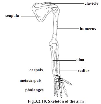
Upper limb or hand - The part of the upper limb from shoulder to the elbow is the arm. It contains one long bone called the humerus. The head of humerus articulates with the glenoid fossa of the scapula. The distal end of the bone articulates with the two forearm bones.
Forearm - This part of the hand is in between the arm and the wrist. The forearm has two bones. They are the ulna and the radius. While the ulna is on the side of the little finger, the radius is on the lateral or thumb side of the forearm.
Wrist - This short region is composed of eight carpal bones. These are arranged into two rows of four each. The carpals along with accompanying ligaments are arranged in such a way that a tunnel on the anterior surface of the wrist called the carpal tunnel has been formed. Tendons, nerves and blood vessels pass through this tunnel to enter the hand.
Hand - The bony framework of the hand is formed of five metacarpals. They are attached to the carpals in the wrist. The concave nature of the palm in the resting position is due to curved arrangement of metacarpals.
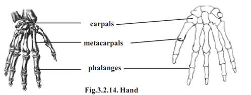
Each hand has five digits. They include one thumb and four fingers. Each digit has small long bones called phalanges. While the thumb has two phalanges other fingers have three each.
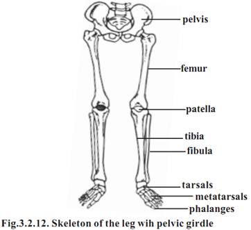
Lower limb or Leg: The general pattern of the lower limb is similar to that of the upper limb.
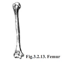
The upper region of the leg is the thigh. It contains a single longest bone called the femur. It has a prominent rounded head for articulating with the acetabulum of the pelvic girdle. The distal end of the femur has two condyles for articulation with the tibia.
The knee region has a large, flat bone called the patella. It articulates with the patellar groove of the femur.
Leg - The leg is that part of the lower limb between the knee and the ankle. It consists of two bones namely, the tibia and the fibula. The tibia is larger and it supports most of the weight of the leg.
Ankle: The ankle consists of seven tarsal bones. The ankle articulates with the tibia and the fibula through the talus.
Foot: It is formed of metatarsals and phalanges. They correspond to the metacarpals and phalanges of the hand.
Joint
All bodily movements are caused by muscles. Our skeletal muscles are firmly attached to bones. Movements involving such muscles cause pull on our bones. Hence movements need movable bone joints.
A joint or an articulation is a place where two bones come together. All joints are not movable. Many joints allow only limited movements.
The joints are named according to the bones that are united.
Kinds of joints - There are three major kinds of joints. They are the fibrous, cartilaginous and synovial joints.
Fibrous joints - In this type, the joints are united by fibrous connective tissue. There is no joint cavity. These joints show little or no movement. Sutures formed between cranial bones, a syndesmosis (to bind) between radius and ulnas are examples for this type.
Cartilaginous joints - These joints unite two bones by means of either hyaline cartilage (synchondroses) or fibrocartilage (symphyses). The articulation between the first rib and the sternum is an example for syncondrosis. Symphysis pubis and intervertebral discs are examples for symphyses.
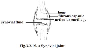
Synovial joints - These joints contain a synovial fluid. This fluid is a complex mixture of polysaccharides, proteins, fats and cells. It forms a thin lubricating film covering the surfaces of a joint. Elbow and knee joints are of this type.
References
T. Sargunam Stephen, Biology (Zoology). First Edition – 2005, Government of Tamilnadu.


|
|
|
|
علامات بسيطة في جسدك قد تنذر بمرض "قاتل"
|
|
|
|
|
|
|
أول صور ثلاثية الأبعاد للغدة الزعترية البشرية
|
|
|
|
|
|
|
مدرسة دار العلم.. صرح علميّ متميز في كربلاء لنشر علوم أهل البيت (عليهم السلام)
|
|
|