Musculature
 المؤلف:
Kelly M. Harrell and Ronald Dudek
 المصدر:
Lippincott Illustrated Reviews: Anatomy
 الجزء والصفحة:
 12-7-2021
 2317
2317
Musculature
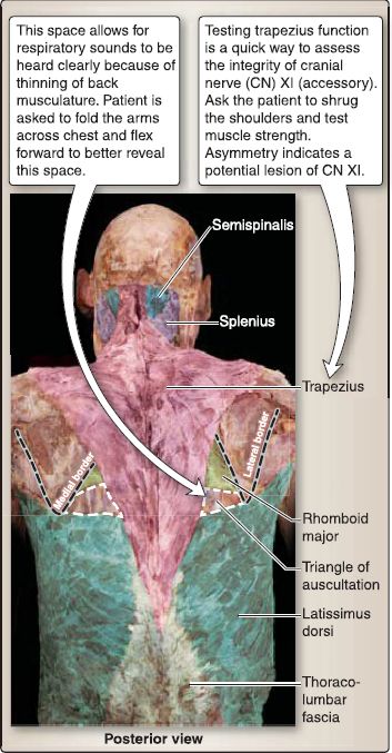
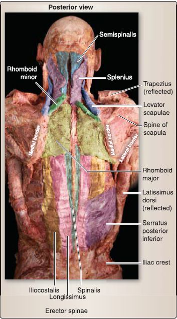
Back musculature can be divided into intrinsic and extrinsic muscle groups, based on developmental origin. Muscles of the back are further arranged into superficial, intermediate, and deep groups (Figs. 1 and 2).

Figure 1: Back musculature.

Figure 2: Superficial, intermediate, and deep back musculature (trapezius and
latissimus dorsi reflected bilaterally).
Extrinsic muscles are found in the superficial and intermediate groups and are innervated by anterior rami, while intrinsic muscles make up the deep group and are innervated by posterior rami. The exception to this is the trapezius, which is innervated by cranial nerve XI, the accessory nerve.
A. Superficial group
Superficial back muscles originate from the spine and insert onto the upper limb . This group comprises two layers: the first layer includes the latissimus dorsi and the trapezius muscles. The second layer includes the rhomboid major and minor and levator scapulae muscles (see Figs. 1and 2).
B. Intermediate group
Intermediate back muscles function to assist respiration and proprioception and to receive segmental innervation (anterior rami) and vascularization. This group includes the serratus posterior superior and inferior muscles (see Fig. 2).
C. Deep group
Deep back muscles are native to the back, meaning during development, they originated posteriorly and remained in this position (Fig. 3; see also Fig. 2). This group is innervated by segmental posterior rami and function bilaterally to maintain an upright, extended spine and unilaterally to side-bend and rotate the spine. This group is further subdivided into superficial, intermediate, and deep layers.

Figure 3 :A, Cervical and thoracic levels showing deeper dissection on the right sides of the cadaver (rhomboid major and minor are reflected on right side). B, Lumbar level computed tomography (Cl) scan showing intrinsic back muscle orientation.
1. Superficial layer: The V-shaped splenius (cervicis and capitis) muscles on the posterior neck resemble a bandage (Latin for bandage). They originate from the midline nuchal ligament and C7-T6 spinous processes and insert onto the mastoid process, superior nuchal line, and the cervical transverse processes (tubercle).
2. Intermediate layer: Erector spinae muscles are a collection of three muscles that originate from a broad tendon arising from the posterior iliac crest, sacrum, sacroiliac and sacrospinous ligaments and lumbar and sacral spinous processes. They run superiorly along the vertebral column. Arranged medially to laterally, these muscles act as the primary extensor of the spine.
a. Spinalis (thoracis, cervicis, and capitis) muscles: These insert onto spinous processes.
b. Longissimus (thoracis, cervicis, and capitis) muscles: These insert onto ribs, transverse processes, and the mastoid process.
c. lliocostalis (lumborum, thoracic, and cervicis) muscles: These insert onto the angles of ribs and cervical transverse processes.
3. Deep layer: The transversospinales muscle group collectively originates from transverse processes and inserts onto spinous processes superiorly. Functionally, these muscles act as spinal stabilizers. This group includes the following muscles, progressing from superficial to deep.
a. Semispinalis (capitis) muscle: This muscle spans five vertebral segments.
b. Multifidus muscle: This muscle spans three vertebral segments.
c. Rotatores muscle: This muscle spans one vertebral segment.
 الاكثر قراءة في علم التشريح
 اخر الاخبار
اخبار العتبة العباسية المقدسة


