Larynx
Multifunctional by design, the larynx plays an important role in both respiration and phonation. It is located in the mid line of the muscular triangle of the neck between the fourth and sixth cervical vertebrae. It is attached superiorly to the hyoid bone by the thyrohyoid membrane and is continuous inferiorly with the trachea .
1. Anatomy: Structurally, the larynx is made up of a cartilaginous scaffold, onto which thick connective tissue membranes and intrinsic laryngeal, infrahyoid, and pharyngeal muscles attach (Fig. 1).
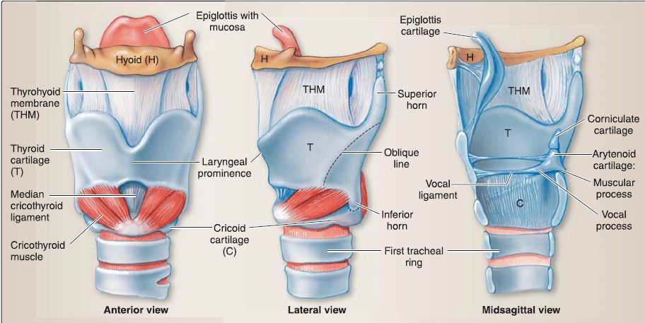
Figure 1: Cartilaginous skeleton of the larynx and associated ligamentous structures.
a. Cartilage: The cartilaginous framework consists of the thyroid, cricoid, arytenoid (2), epiglottic, corniculate (2), and cuneiform (2) cartilages.
[1] Thyroid cartilage: This shield-link cartilage is the largest of the laryngeal cartilages. Right and left lamina fuse anteriorly to form the laryngeal prominence. It is incomplete posteriorly and articulates with the cricoid cartilage inferiorly (Fig. 2).
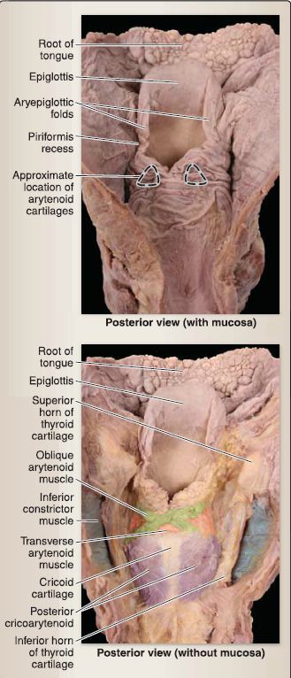
Figure 2: Larynx and laryngopharynx (opened).
[2] Cricoid cartilage: This cartilage is ring shaped and is the most inferior of the thyroid cartilages, located at the C6 vertebral level.
[3] Arytenoids: These paired structures have vocal and muscular processes and articulate on the posterior superior cricoid cartilage. They can abduct, adduct, tilt, and rotate to aid in respiration and phonation.
[4] Epiglottis: This is a singular, leaf-shaped cartilage that borders the laryngeal inlet anteriorly and superiorly and articulates with anterior superior thyroid cartilage. It has a soft tissue muscular fold attached laterally and functions to cover the laryngeal inlet during swallowing. A quadrangular membrane connects the arytenoid cartilages.
b. Ligamentous capsules: The abovementioned cartilaginous articulations possess ligamentous capsules. Additional connections include the cricothyroid ligament, which connects the thyroid and cricoid cartilages anteriorly. The thickened middle portion of the ligament-median cricothyroid ligament-can be palpated easily and is clinically important for securing the airway in an emergency. Vocal ligaments span between the inner surface of the thyroid cartilage and arytenoid vocal process. Superior to the vocal ligaments is the inferior free margin of the quadrangular membrane, which constitutes the vestibular ligaments. The superior free margin constitutes the aryepiglottic ligament.
c. Laryngeal cavity: Between the laryngeal inlet and the inferior border of the cricoid cartilage is the laryngeal cavity. The inner surface of cavity is covered with a thick mucosa. This mucous membrane overlies cartilage, muscles, and ligaments to produce a collection of folds, which serve as boundaries of and within the laryngeal cavity.
[1] Aryepiglottic fold: This mucosa overlies the aryepiglottic ligaments and the distal portion of the oblique arytenoid muscles. It serves as the lateral boundary of the laryngeal inlet.
[2] Vestibular fold: The "false vocal cords" have a protective function. Lying superior to the vocal fold, this mucosa overlies the vestibular ligaments.
[3] Vocal fold: The "true vocal cords" have a phonation function and is mucosa overlying the vocal ligaments and vocalis and thyroarytenoid muscles.
d. Spaces: Spaces within the cavity are named in reference to these folds (Fig. 3). From superior to inferior, these spaces and folds are the laryngeal inlet, laryngeal vestibule, vestibular fold, laryngeal ventricle, vocal fold, and the infraglottic cavity. The spaces located medially between the vestibular and vocal folds are the rima vestibule and rima glottidis, respectively.
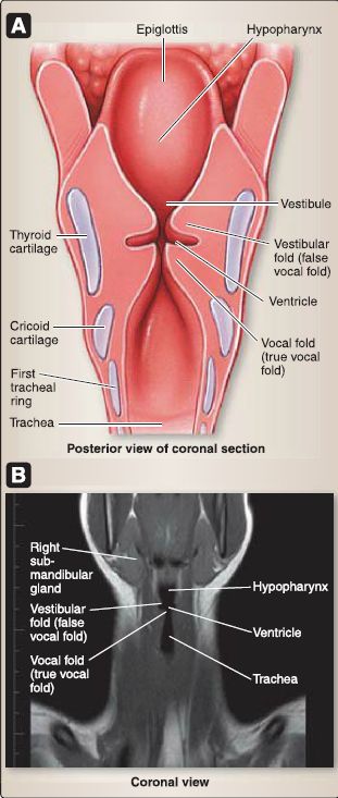
Figure 3: Laryngeal folds and spaces. A, Schematic view. B, Computed tomography scan.
Collectively, the rima glottidis and vocal folds make up the glottis, which constitutes the vocal apparatus.
2. Innervation: General visceral afferent sensation to the laryngeal mucosa is mediated by branches of the vagus nerve (CN X) and divided by at the level of the vocal folds. The internal branch of the superior laryngeal nerve supplies the mucosa above the vocal folds, whereas the inferior laryngeal nerve (continuation of the recurrent laryngeal nerve) supplies the mucosa below the vocal folds.
3. Musculature: Intrinsic muscles of the larynx function to open (abduct) and close (adduct) the airway and alter vocal pitch and tone. All intrinsic laryngeal muscles receive branchiomotor innervation by way of the vagus nerve (CN X).
a. Cricothyroid muscle: Specifically, the cricothyroid muscle is innervated by the external laryngeal branch, and the remaining muscles are innervated by the recurrent laryngeal branch (also referred to as the inferior laryngeal nerve once inside the larynx). Muscle nomenclature describes attachment sites (Figs.3).
b. Vocalis: The vocalis muscle constitutes the medial most fibers of the thyroarytenoid muscle. The vocal is muscle functions to make minute adjustments along the course of the vocal ligament, thus allowing for relaxation of the ligament in one region, while maintaining tension in another (Figs. 4 and 5).
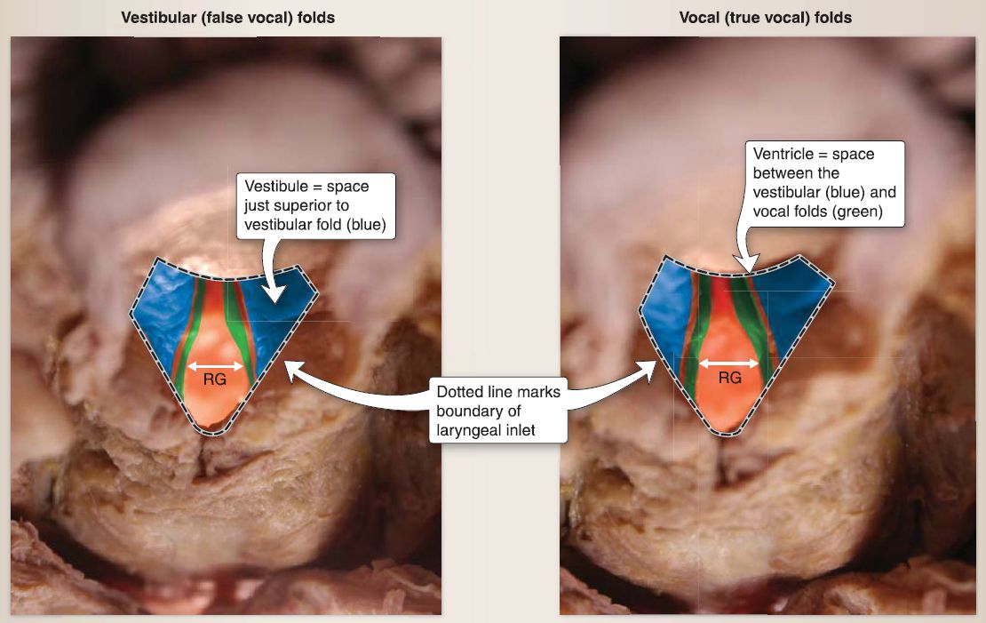 Figure 4: Vocal cords. RG = Rima glottidis
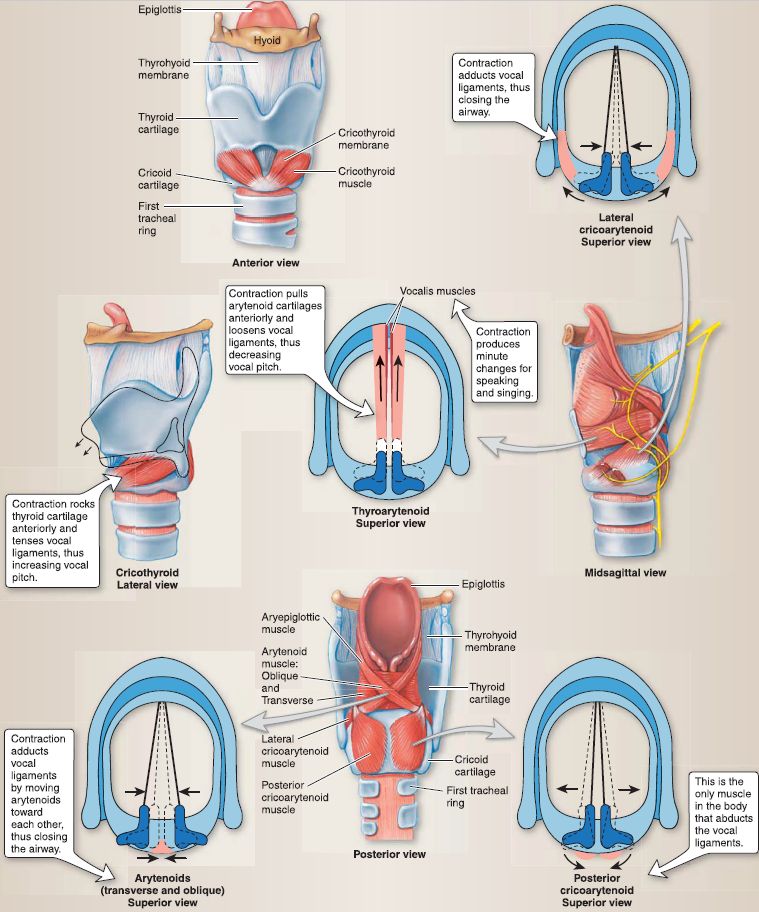
Figure 5: Intrinsic laryngeal structures and muscular actions on vocal ligaments.
4. Vasculature: The larynx receives its blood supply mainly by the superior and inferior laryngeal arteries and branches of the superior and inferior thyroid arteries, respectively. The superior laryngeal artery pierces the thyrohyoid membrane along with the internal laryngeal branch of the superior laryngeal nerve to enter the organ. The inferior laryngeal artery travels superiorly with the recurrent laryngeal nerve on the side of the larynx. Venous drainage occurs by way of veins of the same names.
5. Lymphatics: The lymphatics of the larynx are divided at the level of the vocal folds. Above the vocal folds, lymph vessels follow the superior laryngeal artery to the superior deep cervical nodes. Below the vocal folds, lymph drains into paratracheal and pretracheal nodes before draining into the inferior deep cervical nodes.


