
آخر المواضيع المضافة

 النبات
الحيوان
الأحياء المجهرية
علم الأمراض
التقانة الإحيائية
التقنية الحيوية المكروبية
التقنية الحياتية النانوية
علم الأجنة
الأحياء الجزيئي
علم وظائف الأعضاء
الغدد
المضادات الحيوية
النبات
الحيوان
الأحياء المجهرية
علم الأمراض
التقانة الإحيائية
التقنية الحيوية المكروبية
التقنية الحياتية النانوية
علم الأجنة
الأحياء الجزيئي
علم وظائف الأعضاء
الغدد
المضادات الحيوية| Optical Biosensors |


|
|
Read More
Date: 15-1-2021
Date: 20-1-2021
Date: 22-12-2020
|
Optical Biosensors
Optical biosensors (also called optodes) have generated considerable interest, particularly with respect to the use of fibre optics and optoelectronic transducers. These allow the safe non-electrical remote sensing of materials in hazardous or sensitive (i.e. in vivo) environments. An advantage of optical biosensors is that no reference sensor is needed; a comparative signal is generally easily generated by splitting the light source used by the sampling sensor. A simple example of an optical biosensor is the fibre optic lactate sensor (Figure ) which senses changes in molecular oxygen concentrations by determining its quenching of a fluorescent dye:
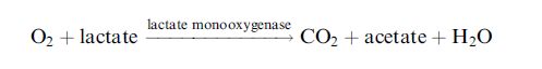
The presence of oxygen quenches (reduces) the amount of fluorescence generated by the dyed film. An increase in lactate concentration reduces the oxygen concentration reaching the dyed film so alleviating the quenching and consequentially causing an increase in the fluorescence output.

Figure . A fibre optic lactate biosensor.
Simple colorimetric changes can be monitored in some biosensor configurations. A lecithin biosensor has been developed containing phospholipase D, choline oxidase and bromothymol blue. The change in pH, due to the formation of the acid betaine from the released choline, causes a change in the bromothymol blue absorbance at 622 nm.22 Gasphase reactions can also be monitored. For example, alcohol vapour can be detected by the colour change of a dry dispersion of alcohol oxidase and peroxidase plus the redox dye 2,6-dichloroindophenol.
One of the most widely established biosensor technologies is the lowtechnology single-use colorimetric assay based on a paper pad impregnated with reagents. This industry revolves mainly round blood and urine analysis with test strips costing only a few cents. A particularly important use for these colorimetric test strips is the detection of glucose.
In this case, the strips contain glucose oxidase and horseradish peroxidase together with a chromogen (e.g. o-toluidine) which changes colour when oxidised by the peroxidase-catalysed reaction with the hydrogen peroxide produced by the aerobic oxidation of glucose:

The colour produced can be evaluated by visual comparison with a test chart or by the use of a portable reflectance meter. Many test strips incorporate anti-interference layers to produce more reproducible and accurate results.
It is possible to link up luminescent reactions to biosensors, as light output is a relatively easy phenomenon to transduce to an electronic output. As an example, the reaction involving immobilised (or free) luciferase can be used to detect the ATP released by the lysis of microorganisms:
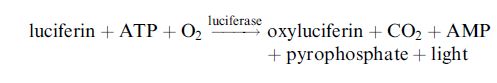
This allows the rapid detection of urinary infections by detecting the microbial content of urine samples.
Evanescent Wave Biosensors
A light beam will be totally reflected when it strikes an interface between two transparent media, from the side with the higher refractive index, at angles of incidence (θ) greater than the critical angle (Figure 1.a).
This is the principle that allows transparent fibres to be used as optical waveguides. At the point of reflection an electromagnetic field is induced which penetrates into the medium with the lower refractive index, usually air or water. This field is called the evanescent wave and it rapidly decays exponentially with the penetration distance and generally has effectively disappeared within a few hundred nanometres. The exact depth of penetration depends on the refractive indices and the wavelength of the light and can be controlled by the angle of incidence. The evanescent wave may interact with the medium and the resultant electromagnetic field may be coupled back into the higher refractive index medium (usually glass) by essentially the reverse process. This gives rise to changes in the light emitted down the waveguide. Thus, it can be used to detect changes occurring in the liquid medium. The necessary surface interactions impose a limitation on the sensitivity of such devices at about 10 pgmm-2 and the requirement to limit non-specific absorption.

Figure . Production of (a) an evanescent wave and (b) surface plasmon resonance. At acute enough angles of incidence the light is totally internally reflected at the glass surface. In (a) an evanescent wave extends from this surface into the air or water medium. This process is amplified in (b) by the presence of the thin metal film.
Various effects, dependent on the biological sensing processes, can be determined, including changes in absorption, optical activity, fluorescence and luminescence. Because of the small degree of penetration, this system is particularly sensitive to biological processes in the immediate vicinity of the surface and independent of any bulk processes or changes.
Due to the small pathlength through the solution, it can even be used for the continuous monitoring of apparently opaque solutions. This biosensor configuration is particularly suitable for immunoassays as there is no need to separate bulk components since the wave only penetrates as far as the antibody–antigen complex. Surface-bound fluorophores may be excited by the evanescent wave and the excited light output detected after it is coupled back into the fibre (Figure 2.).

Figure 2. The principle behind evanescent wave immunosensor. The light output is reduced by absorption within the evanescent wave.
Sensors can be fabricated which measure oxidase substrates using the principle of quenching of fluorescence by molecular oxygen as described earlier. Another advantage of only sensing a surface reaction less than 1 mm thick is that the volume of analyte needed may be very small indeed, that is, less than 1 nl using suitable fluid transfer microfluidics.
Protein A, an important immunoglobulin-binding protein from Staphylococcus aureus, has been determined by this method using a plastic optical fibre coated with its antibody. Detection was by the fluorescence of a fluorescein-bound anti-protein A immunoglobulin which was subsequently bound, sandwiching the protein A.
Surface Plasmon Resonance
The evanescent field generated by the total internal reflection of monochromatic plane-polarised light within a fibre optic or prism may be utilised in a different type of optical biosensor by means of the phenomenon of surface plasmon resonance (SPR). If the surface of the glass is covered with a very thin layer of metal (usually pure gold, silver or palladium, just a nanometre or so thick) then the electrons at the surface may be caused to oscillate in resonance with the photons (as surface plasmon polaritons). This generates a surface plasmon wave and amplifies the evanescent field on the far side of the metal (Figure 1.b). If the metal layer is thin enough to allow penetration of the evanescent field to the opposite surface, the effect is critically dependent on the 100nm or so of medium that is adjacent to the metal.
This effect occurs only when the light is at a specific angle of incidence dependent on its frequency, the thickness of the metal layer and the refractive index of the medium immediately adjacent the metal surface within the evanescent field. The generation of this surface plasmon resonance adsorbs some of the energy of the light so reducing the intensity of the internally reflected light (Figure 3.). Changes occurring in the medium caused by biological interactions may be followed using the consequential changes in the intensity of the reflected light or the resonance angle. Figure 3. shows the change in the resonance angle of a human chorionic gonadotrophin (hCG) biosensor on binding hCG to surface-bound hCG antibody.28 The sensitivity in such devices is limited by the degree of uniformity of the surface and the bound layer and the more-sensitive devices minimise light scattering. Under optimal conditions just 20 or 30 protein molecules bound to each square micrometre of surface may be detected. As with other immunosensors, the main problem occurring in such devices is non-specific absorption.
The biological sensing can be achieved by attaching the bioactive molecule to the medium side of the metal film. Physical adsorption may be used but, because this may lead to undesired denaturation and weak

Figure 3. The change in absorption due to surface plasmon resonance.
binding, covalent binding is often preferred. Gold films can be coated with a monolayer of long-chain 1,o-hydroxyalkylthiols, which are copolymerised to a flexible un-cross-linked carboxymethylated dextran gel, allowing the subsequent binding of bioactive molecules. This flat plate system, marketed as Biacore, allows the detection of parts per million of protein antigen where the appropriate antibody is bound to the gel. Similarly, biosensors for DNA detection can be constructed by attaching a DNA or RNA probe to the metal surface when as little as a few femtograms of complementary DNA or RNA can be detected and, as a bonus, the rate of hybridisation may be determined. Such biosensors retain the advantages of the use of evanescent fields as described earlier. They can also be used to investigate the kinetics of the
binding and dissociation processes. In spite of the relatively low costs possible for producing biosensing surfaces, the present high cost of the instrumentation is restricting developments in this area. However, developments in the use of low-cost light-emitting diodes and lens arrays together with the use of porous surfaces to increase surface area, microfluidics and integrated devices with in-built waveguided interferometry of reference beams are allowing the field to progress rapidly.


|
|
|
|
حمية العقل.. نظام صحي لإطالة شباب دماغك
|
|
|
|
|
|
|
إيرباص تكشف عن نموذج تجريبي من نصف طائرة ونصف هليكوبتر
|
|
|
|
|
|
بمشاركة 60 ألف طالب.. المجمع العلمي يستعدّ لإطلاق مشروع الدورات القرآنية الصيفية
|
|
|
|
صدور العدد الـ 33 من مجلة (الاستغراب) المحكمة
|
|
|
|
المجمع العلمي ينظّم ورشة تطويرية لأساتذة الدورات القرآنية في كربلاء
|
|
|
|
شعبة التوجيه الديني النسوي تختتم دورتها الثانية لتعليم مناسك الحجّ
|