

 النبات
 الحيوان
 الأحياء المجهرية
 علم الأمراض
 التقانة الإحيائية
 التقنية الحيوية المكروبية
 التقنية الحياتية النانوية
 علم الأجنة
 الأحياء الجزيئي
 علم وظائف الأعضاء
 الغدد
الغدد
 المضادات الحيوية
|
Read More
Date: 28-7-2016
Date: 5-1-2017
Date: 11-1-2017
|
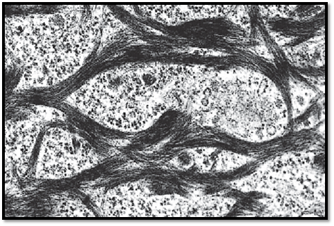
Tonofilaments-Cytokeratin Filaments
Using electron microscopy, the light microscopic images of intracellular tonofibrils prove to be bundles of very fine filaments. The bundles are either strictly parallel or wavy bundles, which create the image of brush strokes in electron micrographs. Tonofibrils pervade especially the cells in the lower layers of the multilayered squamous epithelium. They line up in the direction of the tensile force. However, filament bundles also extend from the cell center to areas with many desmosomes. Tonofilament bundles in the epithelial cell of the vaginal portio uteri.

References
Kuehnel, W.(2003). Color Atlas of Cytology, Histology, and Microscopic Anatomy. 4th edition . Institute of Anatomy Universitätzu Luebeck Luebeck, Germany . Thieme Stuttgar t · New York .


|
|
|
|
دراسة يابانية لتقليل مخاطر أمراض المواليد منخفضي الوزن
|
|
|
|
|
|
|
اكتشاف أكبر مرجان في العالم قبالة سواحل جزر سليمان
|
|
|
|
|
|
|
اتحاد كليات الطب الملكية البريطانية يشيد بالمستوى العلمي لطلبة جامعة العميد وبيئتها التعليمية
|
|
|