آخر المواضيع المضافة

 النبات
الحيوان
الأحياء المجهرية
علم الأمراض
التقانة الإحيائية
التقنية الحيوية المكروبية
التقنية الحياتية النانوية
علم الأجنة
الأحياء الجزيئي
علم وظائف الأعضاء
الغدد
المضادات الحيوية
النبات
الحيوان
الأحياء المجهرية
علم الأمراض
التقانة الإحيائية
التقنية الحيوية المكروبية
التقنية الحياتية النانوية
علم الأجنة
الأحياء الجزيئي
علم وظائف الأعضاء
الغدد
المضادات الحيوية| Blood Smear |


|
|
Read More
Date: 1-8-2016
Date: 27-7-2016
Date: 2-8-2016
|
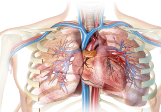
Blood Smear
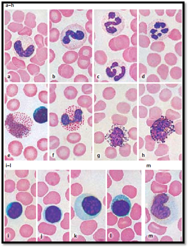
Granulocytes:
a) Neutrophilic granulocyte with banded nucleus.
b) Neutrophilic granulocyte with banded nucleus and beginning segmentation of the nucleus. The cytoplasm is stained light pink and contains azurophilic granules with a diameter of about 0.5 μm.
c) Two neutrophilic granulocytes with segmented nuclei. Fine threads connect the three segments in each of the nuclei. The coarse, heavily stained chromatin bodies are locate d at the peripheral nuclear membrane.
d) Neutrophilic granulocyte, excessively segmented with five segments.
e) Eosinophilic granulocyte (eosinophil) with segmented nucleus. The cell body is filled with coarse eosinophilic (= acidophilic) granules with a diameter of about 1 μm.
f) Eosinophilic granulocyte. The nucleus consists of two round segments.
g) Basophilic granulocyte ( basophil).
h) Basophilic granulocyte. The large nuclei of basophils take up most of the cell body. The cytoplasm contains granules (stained dark violet) of different sizes, which sometimes cover the nucleus.
Lymphocytes:
i) Small lymphocyte. The large eccentric nucleus with large chromatin bodies is lobed and fills the cell body almost completely.
j) Lymphocyte. The large nucleus hardly displays any structural details. It
leaves room for only a small seam of cytoplasm.
k) Large lymphocyte. The eccentric, round nucleus shows coarse bodies. Note the wide seam of cytoplasm.
l) Medium sized lymphocyte with cytoplasmic azurophilic granules.
Monocyte:
m) Monocyte ( mononuclear phagocyte). The nucleus is lobed. Its chromatin has a layered structure. The cytoplasm stains grayish-blue and contains small azurophilic granules.
Stain: Pappenheim (May -Grünwald, Giemsa); magnification: × 900
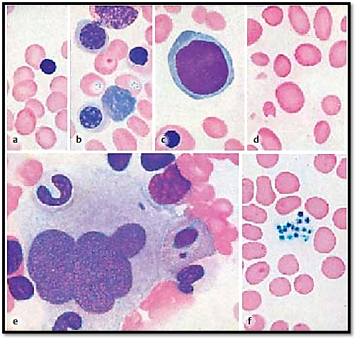
Erythrocytes:
a) Normoblast (orthochromatic erythroblast). Ejected or almost ejected nucleus.
b) At the top of the image, there is a proerythroblast and a macroblast with a polychromatic cell body underneath it. The macroblast nucleus shows a coarse web of chromatin. The normoblast on the right has a pyknotic homogeneous nucleus. Two erythrocytes with basophilic spots are found in the center of the image. The cell underneath is nippe d and damage d. Lower left: macroblast with spoke-like chromatin distribution.
c) Basophilic megaloblast ( promegaloblast) in pernicious anemia. The nucleus is large, is slightly lob e d and features a delicate chromatin web. The cytoplasm (light blue) shows darker blue stained areas in some places. A mature macroblast is locate d at the lower edge of the image.
d) Blood smear with a diagnosis of pernicious anemia. Anisocytosis and poikilocytosis of the erythrocytes.
Megakaryocyte:
e) Mature granular megakaryocyte. Large, lobed, polyploid nucleus. Finely granulated cytoplasm with diffuse border (sternal biopsy material). Megakaryocytes have diameters of more than 50 μm and can be seen at low magnification. Their precursor cells are the megakaryocytes.
Thrombocytes:
f) Thrombocytes and erythrocytes. Thrombocytes are pinched-off cells from megakaryocytes, the giant cells of the bone marrow. A megakaryocyte can produce up to 80 0 0 thrombocytes ( thrombopoiesis).
Stain: Pappenheim (May -Grünwald, Giemsa); magnification: × 900
References
Kuehnel, W.(2003). Color Atlas of Cytology, Histology, and Microscopic Anatomy. 4th edition . Institute of Anatomy Universitätzu Luebeck Luebeck, Germany . Thieme Stuttgart · New York .


|
|
|
|
دراسة يابانية لتقليل مخاطر أمراض المواليد منخفضي الوزن
|
|
|
|
|
|
|
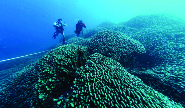
اكتشاف أكبر مرجان في العالم قبالة سواحل جزر سليمان
|
|
|
|
|
|
|
اتحاد كليات الطب الملكية البريطانية يشيد بالمستوى العلمي لطلبة جامعة العميد وبيئتها التعليمية
|
|
|