

 النبات
 الحيوان
 الأحياء المجهرية
 علم الأمراض
 التقانة الإحيائية
 التقنية الحيوية المكروبية
 التقنية الحياتية النانوية
 علم الأجنة
 الأحياء الجزيئي
 علم وظائف الأعضاء
 الغدد
الغدد
 المضادات الحيوية
|
Read More
Date: 2-8-2016
Date: 3-8-2016
Date: 4-1-2017
|
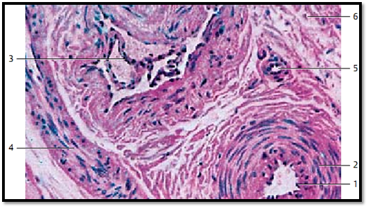
Artery and Vein
This preparation is from the submucosal tissue of the stomach. The micrograph shows cross-sections of a small artery (bottom right) and a vein. The epithelial cell nuclei of the tunica intima protrude into the vascular lumen and make the opening look like an eyelet 1 . Connective tissue dominates in the tunica media of the venous wall. In contrast, in the tunica me dia of the arterial wall circular smooth muscle bundles 2 are more prevalent. Folds of the tunica intima form the venous valves 3 , which extend into the venous lumen.
1 Endothelial cell nuclei
2 Circular musculature
3 Venous valve
4 Arteriole (longitudinal cut)
5 Arteriole (cut across the axis)
6 Connective tissue from the submucous coat (stomach)
Stain: alum hematoxylin-eosin; magnification: × 300

References
Kuehnel, W.(2003). Color Atlas of Cytology, Histology, and Microscopic Anatomy. 4th edition . Institute of Anatomy Universitätzu Luebeck Luebeck, Germany . Thieme Stuttgart · New York .


|
|
|
|
"إنقاص الوزن".. مشروب تقليدي قد يتفوق على حقن "أوزيمبيك"
|
|
|
|
|
|
|
الصين تحقق اختراقا بطائرة مسيرة مزودة بالذكاء الاصطناعي
|
|
|
|
|
|
|
مكتب السيد السيستاني يعزي أهالي الأحساء بوفاة العلامة الشيخ جواد الدندن
|
|
|