آخر المواضيع المضافة

 النبات
الحيوان
الأحياء المجهرية
علم الأمراض
التقانة الإحيائية
التقنية الحيوية المكروبية
التقنية الحياتية النانوية
علم الأجنة
الأحياء الجزيئي
علم وظائف الأعضاء
الغدد
المضادات الحيوية
النبات
الحيوان
الأحياء المجهرية
علم الأمراض
التقانة الإحيائية
التقنية الحيوية المكروبية
التقنية الحياتية النانوية
علم الأجنة
الأحياء الجزيئي
علم وظائف الأعضاء
الغدد
المضادات الحيوية| Antibiotic Resistance |


|
|
Read More
Date: 16-4-2021
Date: 30-10-2020
Date: 8-5-2016
|
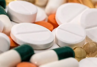
Antibiotic Resistance
In the 1940s, the clinical use of antibiotics first curbed the widespread threat of deadly bacterial infection. These drugs effectively inhibited bacterial growth that had gone unchecked for decades. Antibiotics did not, however, eradicate the threat of bacterial infection. In fact, the widespread use of antibiotics gave a selective advantage to bacteria that had antibiotic resistance. Many strains of bacteria have developed antibiotic resistance, or insensitivity to antibiotic drugs, in response to antibiotic selection pressures. Now, bacteria employ a myriad of resistance mechanisms to circumvent the best efforts of antibiotic researchers and clinicians. Only with the development of new and potent antibiotics and the appropriate use of existing antibiotics will researchers regain control over this resilient lifeform, bacteria.
1. A Historical Perspective
The development of antibiotics as therapeutic agents began in the late 1930s to combat the most common cause of death, infectious disease. In the preantibiotic era, any infection could prove mortal. Subsequently, over 150 different antibiotics have been synthesized or discovered, and these drugs are used to treat bacterial infections, such as pneumonia, malaria, and tuberculosis (1, 2.(
Antibiotics are a collection of natural products and synthetic compounds that kill bacteria. Naturally occurring antibiotics are isolated from molds, yeasts, and bacteria. These organisms use antibiotics as defense mechanisms to kill other bacteria. Alternatively, synthetic antibiotics are developed by understanding the architecture and function of bacteria (see Fig. 1). Some bacteria have cell walls, and many effective antibiotics, such as penicillin, bacitracin, and cephalosporin, inhibit the synthesis of this cell wall. The bacterial machinery for protein biosynthesis differs from that of many host organisms and, therefore, is a good target for antibiotics, such as tetracycline and chloramphenicol. Additionally, antibiotics, such as rifampin and quinolones, specifically inhibit DNA replication in bacteria (1).

Figure 1. Mechanisms of action of some common antibiotics. A schematic of some basic functions of bacterial cells to which antibiotics are targeted. The X indicates an inhibition of that function by the antibiotics noted .
Antibiotics were considered the “wonder drugs” of their time, and in retrospect, this highly favored opinion resulted in their overuse. Antibiotics were commonly prescribed by physicians to cure and to appease their patients. Some patients would have genuine bacterial infections, for which antibiotic treatment is appropriate, whereas others would request antibiotics for viral infections that are not susceptible to these drugs. In addition, individuals who have suppressed immune systems, such as AIDS patients or organ transplants patients, would harbor bacteria that acquire resistance more easily. Antibiotics were also used prophylactically in agriculture and aquaculture industries to keep livestock healthy (3).
By the late 1960s, infectious disease appeared to be under control by the use of a variety of antibiotics. However, antibiotics simply depressed the propagation of bacteria. They did not eradicate it. Nonetheless, research in human health changed its focus from infectious diseases to chronic diseases, and new antibiotics were no longer being developed (2, 4). Many microbiologists warned the human health community that bacteria were potent, infectious pathogens that should not be underestimated. Bacteria have survived for more than three billion years despite numerous environmental changes on earth, and industrial wastes, insecticides, and herbicides. Obviously, the mechanisms bacteria use to survive and adapt to these adversities were very effective.
2. The Origins of Antibiotic Resistance
Even before the first clinical application of antibiotics, antibiotic resistance, the ability of bacteria to evade the deleterious effects of antibiotics, was postulated. In 1940, Abraham and Chain identified a bacterial enzyme that inactivates one of the first antibiotics, penicillin. Then, any bacterium that produces this enzyme would be resistant to penicillin (5). Moreover, microorganisms that use an antibiotic as defense mechanisms would require immunity to that antibiotic. This inherent resistance to a particular antibiotic was defined as a naturally-occurring trait called intrinsic resistance. A few strains of bacteria with intrinsic resistance to a particular antibiotic would not constitute a clinical threat because many diverse antibiotics are available. However, the ability of bacteria to propagate this antibiotic resistance to other strains of bacteria had been underestimated.
The exchange of genetic information between bacteria of the same strain is a common, yet typically slow process. Mechanisms of exchange include (1) conjugation—a single DNA strand from one bacterium enters another bacterium and is replicated as a part of that genome, (2) transduction—foreign DNA is introduced into bacteria by transducing bacteriophages, and (3) transformation—autonomously replicating circular DNA plasmids are obtained by bacteria (1). Originally, it was believed that these genetic exchange mechanisms were restricted to bacteria of the same strain. However, a new method of gene exchange, using integrons, was recently identified (6, 7).
Integrons are independent, mobile elements that encode genes for protein functions, and encode additional DNA to guarantee the integron's expression and integration into the bacterial genome. Integrons effectively generate widespread antibiotic resistance by donating antibiotic resistance genes to any strain of bacteria. Ironically, it is widely believed that integrons evolved only recently in response to antibiotic selection pressure. In other words, the use of antibiotics advanced the widespread occurrence of antibiotic resistance.
In addition to the acquisition of genes by the exchange of genetic information, bacteria also have a high mutation rate that allows them to respond to the selective pressure of antibiotics by using their own genome. For example, if bacteria were subjected to tetracycline, a random mutation in the 30S ribosome to weaken tetracycline binding would be advantageous and, therefore, would be perpetuated by the survival of the tetracycline-resistant bacteria (1). It has also been postulated that housekeeping genes, like acyltransferases, may have mutated to gain the ability to modify and inactivate aminoglycoside antibiotics (8-10).
The widespread phenomenon of antibiotic resistance has developed from the promiscuity of bacteria and their genomes. Initially, intrinsic antibiotic resistances were isolated incidents, but the threat of antibiotics has been readily circumvented using the acquisition of antibiotic resistance genes and the high mutational frequency of individual bacteria.
3. Mechanisms of Antibiotic Resistance
Using both newly acquired genes and their own mutated genes, bacteria utilize three basic mechanisms to support antibiotic resistance. Enzymes that degrade antibiotics inside the cell are key players in antibiotic resistance. Bacteria can also alter their permeability barriers to keep antibiotic concentrations below toxic levels inside the cell. Furthermore, the cellular targets of antibiotics can be modified to evade the effects of the antibiotics. These mechanisms of antibiotic resistance are distinct, but all are obtained through the acquisition or mutation of genes.
3.1. Enzymatic Inactivation of Antibiotics
Degradative enzymes are a common mechanism by which bacteria become resistant to antibiotics. Such enzymes chemically modify antibiotics so that they no longer function. The genes for these degradative enzymes are obtained by acquisition of exogenous genes or mutation of endogenous genes. The expression of these genes also governs the level of antibiotic resistance in bacteria.
b-Lactamases are common examples of degradative enzymes that generate antibiotic resistance. b-Lactamases inactivate b-lactam antibiotics, a structurally similar group of penicillin-like antibiotics, all of which have a b-lactam ring structure. b-Lactamases are typically grouped into two major classes, penicillinases and cephalosporinases, based on their substrate affinity (11, 12). In addition to b-lactamases, other enzymes also degrade different antibiotics, such as the aminoglycosides, gentamycin, tobramycin, and amikacin.
All b-lactam antibiotics function similarly. Their b-lactam ring structure inhibits the final step of bacterial cell wall synthesis. Bacterial cell walls are constructed of alternating N-acetylglucosamine and N-acetyl-muramic acid residues that form long peptidoglycan chains, and the final step in cell wall synthesis involves the enzymatic crosslinking of these peptidoglycan chains by a transpeptidase. Because the b-lactam bond resembles a portion of the peptidoglycan chains, this transpeptidase can mistake a b-lactam antibiotic for its natural substrate and hydrolyze the b-lactam bond. This hydrolysis covalently links the b-lactam drug to the transpeptidase and renders it nonfunctional (13).
Although b-lactamases are effective at degrading some antibiotics, their mere presence is not sufficient to cause clinically relevant antibiotic resistance. In fact, these enzymes are found ubiquitously in almost all bacteria, and in some blue-green algae and mammalian tissues. b-lactamases must be present in sufficient quantities to degrade the b-lactam antibiotics effectively before they inhibit cell wall synthesis. The cellular concentration of a b-lactamase depends on its gene expression, and b-lactam antibiotics are inducers of b-lactamase expression. Furthermore, particular b-lactamases have variable affinities for b-lactam antibiotics. Therefore, the degree of antibiotic resistance due to b-lactamases is based on a combination of the ability of the b-lactam antibiotic to induce the expression of b-lactamase, and its ability to be a substrate for b-lactamase (14).
As researchers began to understand this mechanism of antibiotic resistance, more effective b-lactam antibiotics were developed. The early cephalosporins, like penicillin and amoxicillin, are extremely sensitive to b-lactamases, because these b-lactam antibiotics are potent inducers of b-lactamase expression and good substrates for the b-lactamase. In contrast, the more recently developed antibiotic, imipenem, is a strong inducer of b-lactamase expression but maintains its antibiotic activity because it is a poor substrate for most b-lactamases (15). In addition to these new antibiotics, combination therapies are also being implemented to combat antibiotic resistance. Such therapies are comprised of a b-lactam antibiotic together with b-lactamase inhibitors, like clavulanic acid, sulbactam, and tazobactum (16).
3.2. Altered Permeability Barriers: Pore Proteins and Efflux Systems.
The bacterial cell membrane is the major permeable barrier separating the outside of the cell from the inside. The fluidity of the membrane is generally balanced to include most nutrients, while excluding many toxins. Adjusting this fluidity impedes the function of the membrane. Therefore, bacteria cannot protect themselves by changing the fluidity of their membrane. Instead, bacteria have additional structures that surround the cytoplasmic membrane or form pores through it. The alteration of these structures to exclude antibiotics is another mechanism of antibiotic resistance.
Gram-positive and Gram-Negative Bacteria have distinct structures that surround their cytoplasmic membranes. Most Gram-positive bacteria have thick cell walls that are mechanically quite strong, although very porous. Although the cell wall helps Gram-positive bacteria retain their shape, it does not exclude most antibiotics and, therefore, is not a good barrier. Thus, Gram-positive bacteria are relatively susceptible to the influx of antibiotics. Alternatively, a more effective barrier, a second lipid bilayer or membrane, surrounds Gram-negative bacteria. This outer membrane is partially composed of a lipid, lipopolysaccharide (LPS), that is not commonly found in cytoplasmic membranes. The distinguishing feature of LPS is its decreased fluidity, which makes the LPS bilayer an efficient barrier that prevents the permeation of most hydrophobic antibiotics into Gram-negative bacteria (17).
Enveloped by effective barriers, bacteria use pore-forming proteins, called porins, to obtain nutrients from outside the cell. Porins are transmembrane proteins that function as nonspecific, aqueous channels, and allows nutrients to diffuse across the membrane. Porins generally exclude antibiotics because they are narrow and restrictive. Most antibiotics are large, uncharged molecules that cannot easily traverse the narrow porin channels that are lined with charged amino acid residues. However, some antibiotics enter the bacteria through porins, and the deletion or alteration of these porins to exclude particular antibiotics is linked to antibiotic resistance.
Because bacteria cannot develop barriers that are impermeable to all molecules, some toxins do diffuse into bacteria along with nutrients. Therefore, bacterial cell membranes also contain transport proteins that cross the membranes and use energy to remove toxins. They are called active efflux systems, and some are directly identified as another significant cause of antibiotic resistance.
Many active efflux systems resemble other transport proteins that catalyze the efflux of common, small molecules, like glucose or cations, and it is likely that mutation has modified them to transport antibiotics. Based on their overall structure, mechanism, and sequence homologies, these transport proteins are classified into four families: (1) the major facilitator family; (2) the resistance nodulation division family; (3) the staphylococcal multidrug resistance family; and (4) the ATP-binding cassette) ABC) transporters. Of these four families, only the ABC transporters use the chemical energy generated from the hydrolysis of ATP to drive molecules across the membrane. Members of the three other families use an electrochemical proton gradient, or proton-motive force, as the source of energy (18, 19).
Some active efflux systems exclude a variety of unrelated toxins from the cell. These multidrug resistance (MDR) efflux systems in bacteria are comparable to those found in mammalian cells. An example of a bacterial MDR efflux system is the Bmr transporter that transports drugs which have diverse chemical structures and physical properties and include cationic dyes, rhodamine-6G, ethidium bromide, and the antibiotics netropsin, puromycin, and fluoroquinone (20) . Other MDR efflux systems characterized in bacteria include NorA in Staphylococcus aureus, MexB in Pseudomonas aeruginosa, and EmrB in Escherichia coli. If MDR efflux systems occur extensively in bacteria as the source of many antibiotic resistances, they pose a far more formidable challenge than more specific mechanisms of resistance.
3.3. Modification of the Antibiotic's Target
Antibiotics inhibit bacterial growth by inactivating different key proteins that are essential for bacterial survival (see Fig. 1). However, the antibiotic sensitivity of these target proteins can be altered. Typically, antibiotic targets are altered by reducing their affinity for the antibiotic. Bacteria accomplish this change in affinity several ways. Bacteria acquire exogenous DNA for a mutated target protein that no longer interacts with the antibiotic, yet retains the original target protein's function. Alternatively, bacteria's endogenous genes can be mutated to achieve the same end. In contrast, DNA for novel modifying enzymes can be acquired to alter the antibiotic target posttranslationally, reducing its affinity for the antibiotic.
Altering an antibiotic's target protein directly at the DNA level is a common mechanism of target modification. An example of this modification is the mutation of genes for penicillin-binding proteins (PBPs). PBPs are transpeptidases, previously discussed, that catalyze the final step in bacterial cell wall synthesis. These PBPs have high affinity for penicillin and its derivatives, and the binding of penicillin permanently inactivates PBPs. Originating from both endogenous and exogenous DNA sources, mutated PBPs can have a lower affinity for penicillin. Therefore, PBPs are resistant to the antibiotic, yet still provide a crucial function in bacterial cell wall synthesis (21, 22). Another example of target modification via mutated DNA is a single amino acid mutation in the quinolone resistance-determining region of the DNA gyrase gene, gyrA, that can provide up to a 20-fold increase in quinolone resistance (23).
In addition to using mutated antibiotic targets, bacteria can acquire new genes that produce proteins that, in turn, alter antibiotic targets. A well-studied example is the resistance of Staphylococci to erythromycin. It is known that Staphylococci have acquired genes to produce a protein that methylates a residue on the 23S ribosome. The 23S ribosome is the target of erythromycin, but methylated 23S ribosome has a low affinity for erythromycin. This exogenous gene is expressed and prevents the binding of erythromycin to the ribosomes, making the bacteria erythromycin-resistant (24).
References
1. H. C. Neu (1992) Science 257, 1064–1073.
2. J. Travis (1994) Science 264, 360–362.
3. M. Castiglia and R. A. J. Smego (1997) J. Am. Pharm. Assoc. NS37, 383–387.
4. G. H. Cassell (1997) FEMS Immunol. Med. Microbiol. 18, 271–274.
5. E. P. Abraham and E. Chain (1940) Nature 146, 837.
6. H. W. Stokes and R. M. Hall (1989) Mol. Microbiol. 3, 1669–1683.
7. C. M. Collis, G. Grammaticopoulos, J. Briton, H. W. Stokes, and R. M. Hall (1993) Mol. Microbiol. 9, 41–52.
8. T. Udou, Y. Mizuguchi, and R. J. J. Wallace (1989) FEMS Microbiol. Lett. 48, 227–230.
9. K. J. Shaw et al. (1992) Antimicrob. Agents Chemother. 36, 1447–1455.
10. P. N. Rather, E. Orosz, K. J. Shaw, R. Hare, and G. Miller (1993) J. Bacteriol. 175, 6492–6498.
11. M. H. Richmond and R. B. Sykes (1973) Adv. Microb. Physiol. 9, 31–88.
12. K. Bush (1989) Antimicrob. Agents Chemother. 33, 259–276.
13. A. Tomasz (1979) Annu. Rev. Microbiol. 33, 113–137.
14. D. M. Livermore (1993) J. Antimicrob. Chemother. 31 (suppl. A), 9–21.
15. J. Y. Jacobs, D. M. Livermore, and K. W. M. Davy (1984) J. Antimicrob. Chemother. 14, 221229- .
16. K. Coleman et al. (1994) J. Antimicrob. Chemother. 33, 1091–1116.
17. P. R. Cullis and M. J. Hope (1985) In Biochemistry of Lipids and Membranes (D. E. Vance and J. E. Vance, eds.), Benjamin and Cummings, New York, Chap. "2".
18. S. B. Levy (1992) Antimicrob. Agents Chemother. 36, 695–703.
19. K. Lewis, D. C. Hooper, and M. Ouellette (1997) ASM News 63, 605–610.
20. A. A. Neyfakh, V. E. Bidnenko, and L. B. Chen (1991) Proc. Natl. Acad. Sci. USA 88, 47814785- .
21. B. G. Spratt and K. D. Cromie (1988) Rev. Infect. Dis. 10, 699–711.
22. J. M. Ghuysen (1991) Annu. Rev. Microbiol. 45, 37–67.
23. G. A. Jacoby and A. A. Medeiros (1991) Antimicrob. Agents Chemother. 35, 1697–1704.
24. R. Leclercq and P. Courvalin (1991) Antimicrob. Agents Chemother. 35, 1267–1272.


|
|
|
|
"عادة ليلية" قد تكون المفتاح للوقاية من الخرف
|
|
|
|
|
|
|
ممتص الصدمات: طريقة عمله وأهميته وأبرز علامات تلفه
|
|
|
|
|
|
|
ضمن أسبوع الإرشاد النفسي.. جامعة العميد تُقيم أنشطةً ثقافية وتطويرية لطلبتها
|
|
|