آخر المواضيع المضافة

 النبات
الحيوان
الأحياء المجهرية
علم الأمراض
التقانة الإحيائية
التقنية الحيوية المكروبية
التقنية الحياتية النانوية
علم الأجنة
الأحياء الجزيئي
علم وظائف الأعضاء
الغدد
المضادات الحيوية
النبات
الحيوان
الأحياء المجهرية
علم الأمراض
التقانة الإحيائية
التقنية الحيوية المكروبية
التقنية الحياتية النانوية
علم الأجنة
الأحياء الجزيئي
علم وظائف الأعضاء
الغدد
المضادات الحيوية| Bacillus anthracis |


|
|
Read More
Date: 14-3-2016
Date: 15-3-2016
Date: 10-3-2016
|
Anthrax is a rare disease in the United States. For example, from 1984 to 1997, only three case of cutaneous anthrax were reported.
However, in 2001, 20 new cases occurred—11 cutaneous and 11 inhalation anthrax. These infections resulted from probable expo sure to B. anthracis powder sent through the mail.
1. Epidemiology:
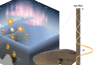
Anthrax is an enzootic disease of worldwide occurrence. [Note: The term enzootic disease applies to a population of animals (equivalent to endemic disease in a human population, that is, its occurrence changes little over time). This is as compared to an epizootic disease, which attacks a large number of animals at the same time (similar to a human epidemic).] Anthrax affects principally domestic herbivores (for example, sheep, goats, and horses) and is transmitted to humans by contact with infected animal products or contaminated dust (Figure 1). Infection is usually initiated by the subcutaneous inoculation of spores through incidental skin abrasions. Less frequently, the inhalation of spore-laden dust causes a pulmonary form of anthrax. [Note: Sometimes an occupational hazard, this form of pneumonia is known as “woolsorter’s disease.”] B. anthracis spores may remain viable for many years in contaminated pastures and in bones, wool, hair, hides, and other animal materials. These spores, like those of clostridia, are highly resistant to physical and chemical agents. In the United States, a veterinary vaccine in widespread use makes domestic animal sources of the disease quite rare. Contaminated agricultural imports may account for the few cases seen and lead occasionally to the quarantine of goods from endemic areas. B. anthracis is a potential bioterrorism agent because it can be easily grown in large quantities. Moreover, the spores are resistant to destruction and can be formulated into an aerosol for wide dissemination. Physicians must be prepared to recognized anthrax even though it is rarely seen in the United States.

Fig1. Anthrax in animal and human hosts.
2. Pathogenesis:
B. anthracis produces a unique capsule that is comprised of poly-D-glutamic acid and is anti-phagocytic. Elaboration of this capsule is essential for full virulence. The organism also produces two plasmid-coded exotoxins: edema toxin and lethal toxin. Both toxins are AB type toxins with activity and binding domains. The binding subunit shared by both toxins is called protective antigen (so named because of its use in producing protective anthrax vaccines). This domain mediates cell entry of both toxins. The activity subunits are called edema factor and lethal factor. Edema factor is a calmodulin-dependent adenylyl cyclase, which causes elevation of intracellular cAMP, resulting in the severe edema usually seen in B. anthracis infections. Lethal factor is responsible for tissue necrosis. Lethal factor complexed with protective antigen is known as lethal toxin, whereas edema factor complexed with protective antigen is known as edema toxin .
3. Clinical significance
a. Cutaneous anthrax: About 95 percent of human cases of anthrax are cutaneous. Upon introduction of organisms or spores that germinate, a papule develops. It rapidly evolves into a painless, black, severely swollen “malignant pustule,” which eventually crusts over. The organisms may invade regional lymph nodes and then the general circulation, leading to fatal septicemia. Although some cases remain localized and heal, the overall mortality in untreated cutaneous anthrax is about 20 percent.
b. Pulmonary anthrax (woolsorter’s disease): Caused by inhalation of spores, the pulmonary form is characterized by progressive hemorrhagic lymphadenitis (inflammation of the lymph nodes), hemorrhagic mediastinitis (inflammation of the mediastinum) and has a mortality rate approaching 100 percent if left untreated.
4. Laboratory identification:
B. anthracis is easily recovered from clinical materials, where it is often present in massive numbers. Microscopically, the organisms appear as blunt-ended bacilli that occur singly; in pairs; or frequently in long chains (Figure 2). They do not sporulate often in clinical samples but do so in culture. The spores are oval and centrally located. On blood agar, the colonies are large, grayish, and nonhemolytic, with an irregular border. Unlike many bacillus species, B. anthracis is nonmotile and is encapsulated in vivo. A direct immunofluorescence assay aids in identification of the organism.

Fig2. Summary of anthrax disease. 1 Indicates first-line drugs.
5. Treatment:
B. anthracis is sensitive to a variety of antibiotics. Cutaneous anthrax responds to ciprofloxacin (see Figure 2). Penicillin is not recommended because of inducible β-lactamase in B. anthracis. Multidrug therapy (for example, ciprofloxacin plus rifampin plus vancomycin) is recommended for inhalation anthrax. Aggressive therapy is indicated for inhalation anthrax both because of the severity of the disease and the fact that the disease is often not diagnosed until late in the course of the illness.
6. Prevention:
A cell-free vaccine is available for workers in high-risk occupations . Postexposure prophylaxis with ciprofloxacin or doxycycline is recommended. [Note: Because of the resistance of endospores to chemical disinfectants, autoclaving is the most reliable means of decontamination.]


|
|
|
|
كيف تجعلين طفلك ينام بسلام طوال الليل؟
|
|
|
|
|
|
|
دراسة: الذكاء الاصطناعي يتفوق على البشر في مراقبة القلب
|
|
|
|
|
|
|
الأمانة العامة للعتبة الكاظمية المقدسة تسجل مشاركتها في مهرجان عين الحياة
|
|
|