


النبات

مواضيع عامة في علم النبات
الجذور - السيقان - الأوراق
النباتات الوعائية واللاوعائية
البذور (مغطاة البذور - عاريات البذور)
الطحالب
النباتات الطبية
الحيوان
مواضيع عامة في علم الحيوان
علم التشريح
التنوع الإحيائي
البايلوجيا الخلوية
الأحياء المجهرية
البكتيريا
الفطريات
الطفيليات
الفايروسات
علم الأمراض
الاورام
الامراض الوراثية
الامراض المناعية
الامراض المدارية
اضطرابات الدورة الدموية
مواضيع عامة في علم الامراض
الحشرات
التقانة الإحيائية
مواضيع عامة في التقانة الإحيائية
التقنية الحيوية المكروبية
التقنية الحيوية والميكروبات
الفعاليات الحيوية
وراثة الاحياء المجهرية
تصنيف الاحياء المجهرية
الاحياء المجهرية في الطبيعة
أيض الاجهاد
التقنية الحيوية والبيئة
التقنية الحيوية والطب
التقنية الحيوية والزراعة
التقنية الحيوية والصناعة
التقنية الحيوية والطاقة
البحار والطحالب الصغيرة
عزل البروتين
هندسة الجينات
التقنية الحياتية النانوية
مفاهيم التقنية الحيوية النانوية
التراكيب النانوية والمجاهر المستخدمة في رؤيتها
تصنيع وتخليق المواد النانوية
تطبيقات التقنية النانوية والحيوية النانوية
الرقائق والمتحسسات الحيوية
المصفوفات المجهرية وحاسوب الدنا
اللقاحات
البيئة والتلوث
علم الأجنة
اعضاء التكاثر وتشكل الاعراس
الاخصاب
التشطر
العصيبة وتشكل الجسيدات
تشكل اللواحق الجنينية
تكون المعيدة وظهور الطبقات الجنينية
مقدمة لعلم الاجنة
الأحياء الجزيئي
مواضيع عامة في الاحياء الجزيئي
علم وظائف الأعضاء
الغدد
مواضيع عامة في الغدد
الغدد الصم و هرموناتها
الجسم تحت السريري
الغدة النخامية
الغدة الكظرية
الغدة التناسلية
الغدة الدرقية والجار الدرقية
الغدة البنكرياسية
الغدة الصنوبرية
مواضيع عامة في علم وظائف الاعضاء
الخلية الحيوانية
الجهاز العصبي
أعضاء الحس
الجهاز العضلي
السوائل الجسمية
الجهاز الدوري والليمف
الجهاز التنفسي
الجهاز الهضمي
الجهاز البولي
المضادات الميكروبية
مواضيع عامة في المضادات الميكروبية
مضادات البكتيريا
مضادات الفطريات
مضادات الطفيليات
مضادات الفايروسات
علم الخلية
الوراثة
الأحياء العامة
المناعة
التحليلات المرضية
الكيمياء الحيوية
مواضيع متنوعة أخرى
الانزيمات

Osteology of Upper Limb
 المؤلف:
Kelly M. Harrell and Ronald Dudek
المؤلف:
Kelly M. Harrell and Ronald Dudek
 المصدر:
Lippincott Illustrated Reviews: Anatomy
المصدر:
Lippincott Illustrated Reviews: Anatomy
 الجزء والصفحة:
 21-7-2021
 7938
7938


+
-
20
Osteology of Upper Limb
The upper limbs are designed for mobility and manipulation (completion of a task). The upper limb is divided into shoulder, arm, forearm, and hand regions. The arrangement of joints and increased range of motion of the upper limb allow for bilateral coordination of crude and fine motor skills. Fine motor skills are achieved directly through the hand and fingers, but range of motion and strength at the more proximal joints are necessary for proper positioning of the hand.
Proximally, the shoulder (pectoral) girdle-comprising the manubrium of the sternum, the clavicles, and the scapulae-forms an incomplete bony ring that connects the upper limb to the axial skeleton. Posteriorly, the bony ring is supported by muscular attachments between the scapula and vertebral column.
Bones of the upper limb (proximal to distal) include the clavicle, scapula, humerus, radius, ulna, carpal bones, metacarpals, and phalanges.
A. Clavicle
The clavicle is an S-shaped bone that acts as a strut to connect the upper limb to the axial skeleton (Fig.1). Medially, the clavicle is convex anteriorly and articulates with the manubrium (sternal end). Laterally, the clavicle is concave anteriorly and flattens to articulate with the acromion process (acromial end). The rough inferior surface of the clavicle has raised areas for ligament and muscle attachment, including the impression for the costoclavicular ligament, groove for the subclavius muscle, and trapezoid line and conoid tubercle for the two parts of the coracoclavicular ligament.
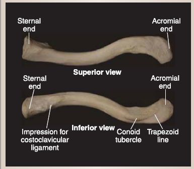
Figure 1: Clavicular osteology.
B. Scapula
The scapula is a triangular bone characterized by medial, lateral, and superior borders and superior and inferior angles (Fig. 2). Scapular structures are often best appreciated from lateral, posterior, and anterior views.
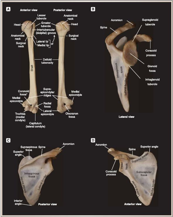
Figure 2: Humeral and scapular osteology. A. Humerus. B-D. Scapula. Hole is an artifact in the bone (*).
1. Lateral: The glenoid fossa serves as the shallow socket portion of the glenohumeral (shoulder) joint, which articulates with the head of the humerus laterally. Supraglenoid and infraglenoid tubercles are located superior and inferior to the fossa, respectively.
2. Posterior: The scapula is unevenly divided posteriorly into supraspinous and infraspinous fossae by a ridge of bone called the spine of the scapula. The spine extends laterally and ends at the acromion process (point of the shoulder).
3. Anterior: The scapula has a concave anterior surface called the subscapular fossa. Arising from the neck of the scapula of the anterior superior surface is the beak-like coracoid process. The suprascapular notch lies medial to the root of the coracoid process.
C. Humerus
The humerus (arm; brachium) is the largest bone of the upper limb (see Fig. .2). Humeral structures can be described from proximal to distal.
1. Proximal: The proximal humerus is characterized by a round head (ball portion of the shoulder joint) that is covered with articular cartilage. The anatomical neck lies between the head and greater and lesser tubercles-two bony prominences that serve as muscle attachment sites (Fig. 3). The greater tubercle is positioned more superolaterally, whereas the lesser is more inferomedially. The intertubercular groove (sulcus; bicipital groove) lies between the tubercles. Extending inferiorly from the greater and lesser tubercle are the lateral and medial lips of the intertubercular groove, respectively. The surgical neck is found just distal to the tubercles.
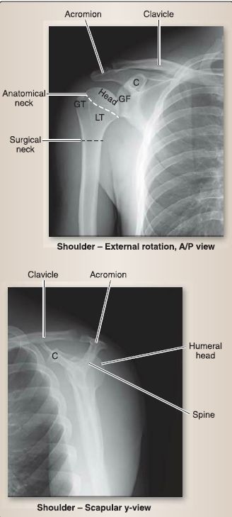
Figure 3: Plain film radiographs of shoulder complex. C = coracoid process, GF = glenoid fossa, GT = greater tubercle, LT = lesser tubercle.
2. Shaft: The humeral shaft is primarily smooth, except for a small midshaft deltoid tubercle laterally and a spiral (radial) line posteriorly.
3. Distal: The distal humerus has a spool-shaped medial condyle (trochlea) and a rounded lateral condyle (capitulum) that articulate with the ulna and radius, respectively. Superior to the condyles are the medial and lateral epicondyles. The coronoid and radial fossae are located between the epicondyles anteriorly and the olecranon fossa posteriorly.
D. Ulna and radius
The bones of the forearm (antebrachium) include the ulna medially and the radius laterally (Figs. 4 and 5). These bones articulate proximally at the elbow with the humerus and distally at the wrist with the carpal bones. They are connected along their length by an interosseous membrane.
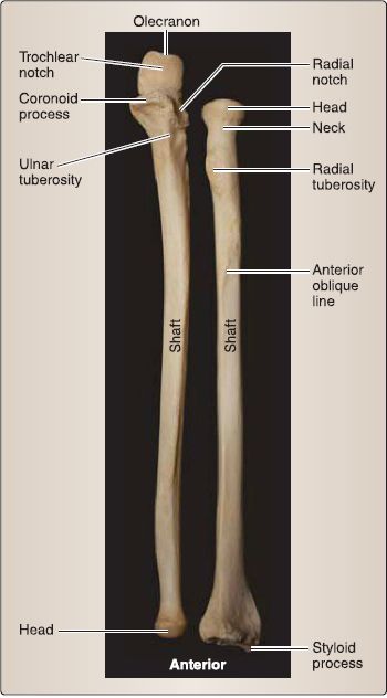
Figure 4: Forearm osteology {ulna and radius).
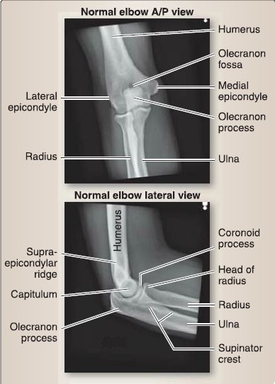
Figure 5: Plain film radiographs of the elbow.
1. Ulnar features: The ulna is characterized proximally by a large trochlear notch framed anteriorly by the coronoid process and posteriorly by the olecranon process, a shallow radial notch, and a raised ulnar tuberosity. The trochlear notch articulates with the trochlea of the humerus, while the radial notch articulates with the head of the radius. Distally, the ulna tapers into a head and styloid process.
2. Radial features: The radius is characterized proximally by a head, neck, and raised radial tuberosity. The head articulates with the capitulum of the humerus. Distally, the radius expands and possesses a styloid process.
E. Carpal and metacarpal bones and phalanges
The carpal and metacarpal bones make up the bony structure of the hand (palm and dorsum), while the phalanges form digits one through five (Fig. 6). Carpal bones are arranged into proximal and distal rows, with bones of the proximal row contributing to the wrist (radiocarpal) joint.
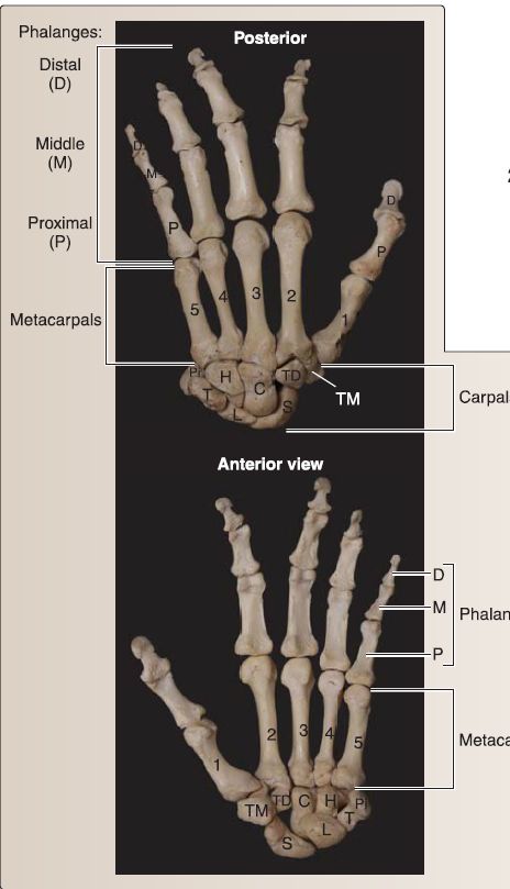
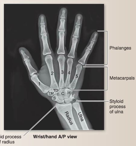
Figure 6: Osteology of the hand with plain film radiograph. C = capitate, H = hamate, L = lunate, Pi = pisiform, S = scaphoid, T = triquetrum, TD = trapezoid, TM = trapezium.
1. Carpal bones: The carpal bones are arranged in a proximal and a distal row.
a. Proximal row: From lateral to medial (radial to ulnar), the proximal row consists of the scaphoid, lunate, triquetrum, and pisiform. The scaphoid, lunate, and triquetrum articulate with the distal radius to form the wrist joint.
b. Distal row: The distal row consists of the trapezium, trapezoid, capitate, and hamate. The distal row articulates with the bases of the five metacarpal bones.
2. Metacarpals: Metacarpals have a proximal base, middle shaft, and distal head. Metacarpal heads articulate with the base of the proximal phalanges.
3. Phalanges: Proximal, middle, and distal phalanges are found in digits two through five, while the first digit (thumb; pollex) only has a proximal and distal phalanx.
F. Surface anatomy
Several surface landmarks of the upper limb are important during physical examination (Fig. 7). Assessing symmetry of the upper limb, from the shoulder to the hand, is essential when formulating a diagnosis. Symmetry and level of these landmarks can be assessed in sitting, standing, or supine positions to determine where along the upper limb deviation may be occurring. Palpating soft-tissue structures, such as tendons, is made easier when patient is asked to actively engage the targeted muscle.
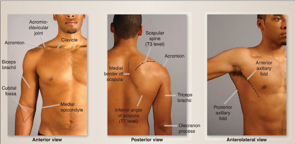
Figure 7: Surface landmarks of the upper limb.
1. Scapula: Posteriorly, the medial and inferior border of the scapula can be palpated as well as the spine (T3 level), superior angle (f 2 level), and inferior angle (f 7 level). Follow the spine out laterally to the point of the shoulder where it becomes the acromion. The articulation between the distal clavicle and acromion (acromioclavicular joint) can be palpated anteriorly and the coracoid process just inferomedial to the acromioclavicular joint.
2. Axillary folds: The anterior axillary fold is made up of the lateral border of the pectoralis major muscle, while the posterior axillary fold is made up of the lateral borders of latissimus dorsi and teres major muscles. These folds provide anterior and posterior boundaries for the axilla, respectively.
3. Proximal and distal bony landmarks: Proximal humeral bony landmarks include the greater tubercle found just inferolateral to the acromion. Moving medially, the intertubercular groove can be palpated and the lesser tubercle most medially. The long head of the biceps brachii tendon lies in the intertubercular groove. Distally, the medial and lateral epicondyles are easily palpated and serve as common tendon attachment sites for the forearm flexors and extensors, respectively. At the elbow, the olecranon process is easily palpated posteriorly, but the other proximal ulnar structures may be difficult to identify. With passive or active pronation/supination, the radial head can be located just distal to the lateral epicondyle.
4. Cubital fossa (anterior elbow): The biceps brachii tendon can be palpated easily with active elbow flexion. The brachia! artery pulse can be found just medial to the biceps tendon. Superficially, the median cubital vein may be visible through the skin (site for venous puncture).
5. Wrist: Both ulnar and radial styloid processes can be palpated with abduction and adduction of the hand, respectively. The radial artery pulse is found just lateral to the flexor carpi radialis tendon and the wrist. On the dorsum of the hand, the anatomical snuff box boundaries can be easily visualized with active thumb abduction and extension. The scaphoid bone and radial styloid process can be palpated within this space. In the hand, the thenar and hypothenar eminences represent the underlying intrinsic thumb and fifth digit ("pinky") muscles, respectively.
 الاكثر قراءة في علم التشريح
 اخر الاخبار
اخبار العتبة العباسية المقدسة

الآخبار الصحية


 قسم الشؤون الفكرية يصدر كتاباً يوثق تاريخ السدانة في العتبة العباسية المقدسة
 "المهمة".. إصدار قصصي يوثّق القصص الفائزة في مسابقة فتوى الدفاع المقدسة للقصة القصيرة
 (نوافذ).. إصدار أدبي يوثق القصص الفائزة في مسابقة الإمام العسكري (عليه السلام)


