
النبات

مواضيع عامة في علم النبات

الجذور - السيقان - الأوراق

النباتات الوعائية واللاوعائية

البذور (مغطاة البذور - عاريات البذور)

الطحالب

النباتات الطبية


الحيوان

مواضيع عامة في علم الحيوان

علم التشريح

التنوع الإحيائي

البايلوجيا الخلوية


الأحياء المجهرية

البكتيريا

الفطريات

الطفيليات

الفايروسات


علم الأمراض

الاورام

الامراض الوراثية

الامراض المناعية

الامراض المدارية

اضطرابات الدورة الدموية

مواضيع عامة في علم الامراض

الحشرات


التقانة الإحيائية

مواضيع عامة في التقانة الإحيائية


التقنية الحيوية المكروبية

التقنية الحيوية والميكروبات

الفعاليات الحيوية

وراثة الاحياء المجهرية

تصنيف الاحياء المجهرية

الاحياء المجهرية في الطبيعة

أيض الاجهاد

التقنية الحيوية والبيئة

التقنية الحيوية والطب

التقنية الحيوية والزراعة

التقنية الحيوية والصناعة

التقنية الحيوية والطاقة

البحار والطحالب الصغيرة

عزل البروتين

هندسة الجينات


التقنية الحياتية النانوية

مفاهيم التقنية الحيوية النانوية

التراكيب النانوية والمجاهر المستخدمة في رؤيتها

تصنيع وتخليق المواد النانوية

تطبيقات التقنية النانوية والحيوية النانوية

الرقائق والمتحسسات الحيوية

المصفوفات المجهرية وحاسوب الدنا

اللقاحات

البيئة والتلوث


علم الأجنة

اعضاء التكاثر وتشكل الاعراس

الاخصاب

التشطر

العصيبة وتشكل الجسيدات

تشكل اللواحق الجنينية

تكون المعيدة وظهور الطبقات الجنينية

مقدمة لعلم الاجنة


الأحياء الجزيئي

مواضيع عامة في الاحياء الجزيئي


علم وظائف الأعضاء


الغدد

مواضيع عامة في الغدد

الغدد الصم و هرموناتها

الجسم تحت السريري

الغدة النخامية

الغدة الكظرية

الغدة التناسلية

الغدة الدرقية والجار الدرقية

الغدة البنكرياسية

الغدة الصنوبرية

مواضيع عامة في علم وظائف الاعضاء

الخلية الحيوانية

الجهاز العصبي

أعضاء الحس

الجهاز العضلي

السوائل الجسمية

الجهاز الدوري والليمف

الجهاز التنفسي

الجهاز الهضمي

الجهاز البولي


المضادات الميكروبية

مواضيع عامة في المضادات الميكروبية

مضادات البكتيريا

مضادات الفطريات

مضادات الطفيليات

مضادات الفايروسات

علم الخلية

الوراثة

الأحياء العامة

المناعة

التحليلات المرضية

الكيمياء الحيوية

مواضيع متنوعة أخرى

الانزيمات
Lymphatic System
المؤلف:
Kelly M. Harrell and Ronald Dudek
المصدر:
Lippincott Illustrated Reviews: Anatomy
الجزء والصفحة:
9-7-2021
3353
Lymphatic System
The lymphatic system comprises an overflow system that drains surplus extracellular tissue fluid and leaked plasma proteins and returns them back into the venous bloodstream. In addition, the lymphatic system functions in the absorption and transport of dietary fat along with immune defense. All regions of the body have lymphatic drainage, even the dural sinuses of the brain. The lymphatic system consists of lymphatic capillaries, superficial and deep lymphatic vessels, lymphatic trunks, right lymphatic duct, thoracic duct, and superficial and deep lymph nodes (Fig. 1).

Figure 1 : Lymphatic system.
A. Lymph vessels
The lymphatic capillaries begin blindly in the extracellular spaces of tissues and form a lymphatic plexus intertwined with a capillary plexus. The lymphatic vessels in general are a bodywide network of vessels with closely spaced valves and are found nearly everywhere blood capillaries are present. The superficial lymphatic vessels follow the venous drainage and eventually drain into the deep lymphatic vessels.
The lymph fluid in the superficial and deep lymphatic vessels passes through several sets of series of superficial or deep lymph nodes as it courses proximally. Large lymphatic vessels converge to form lymphatic trunks.
B. Lymph ducts
The lymphatic trunks unite to form the right lymphatic duct and the thoracic duct (see Fig. 1).
1. Right lymphatic duct: This duct begins by the union of the right subclavian, right jugular, and right bronchomediastinal lymph trunks. It terminates at the junction of the right internal jugular vein and right subclavian vein (i.e., right brachiocephalic vein) at the base of the neck. The right lymphatic duct drains lymph from the 1) right side of the head and neck, 2) right breast (medial and lateral quadrant), 3) right upper limb/superficial thoracoabdominal wall, and 4) right lung.
2. Thoracic duct: This duct begins in a majority of individuals as the abdominal confluence of lymph trunks at the L 1/L2 vertebral level. The confluence of lymph trunks receives lymph from four main lymphatic trunks: the right and left lumbar lymph trunks and the right and left intestinal lymph trunks. In a small percentage of individuals, the abdominal confluence of lymph trunks is represented as a dilated sac (cisterna chyli). The thoracic duct terminates at the junction of the left internal jugular vein and left subclavian vein (i.e., left brachiocephalic vein) at the base of the neck. The thoracic duct drains lymph from 1) the left side of the head and neck, 2) left breast, 3) left upper limb/superficial thoraco-abdominal wall, and 4) all the body below the diaphragm.
C. Lymph nodes
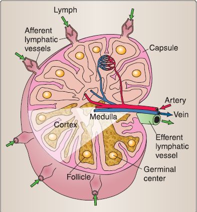
The lymph nodes are bean-shaped glands that lie in the course of the lymphatic vessels (see Fig. 1). They filter and monitor lymph for various antigens.
1. Layers: The lymph node consists of the outer cortex, inner cortex, and medulla (Fig. 2). The outer cortex contains a number of cell types: mature (or virgin) lgM+ and lgD+ B lymphocytes, follicular dendritic cells, macrophages, and fibroblasts (or reticular cells). The inner cortex differs in that, instead of B lymphocytes, it contains mature T lymphocytes. The medulla contains all of the cell types contained in the outer and inner cortices as well as plasma cells.

Figure 2 : Lymph node organization.
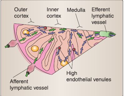
2. Lymphatic drainage: Afferent lymphatic vessels carry lymph, which enters the lymph node on the convex surface (Fig. 3). The lymph percolates through the subcapsular, trabecular, and finally the medullary sinuses. The medullary sinuses coalesce into the efferent lymphatic vessel that exits at the hilus. A majority of B lymphocytes and T lymphocytes exit the lymph node via the efferent lymphatic vessel.

Figure 3 :Lymph drainage through a lymph node.
3. Blood supply: Arterioles enter the lymph node at the hilus, give off some branches in the medulla, and then travel to the cortex where they form a capillary network. The capillaries drain into postcapillary venules (also called high endothelial venules) within the inner cortex where a majority of the B lymphocytes and T lymphocytes enter the lymph node via the postcapillary venules. The postcapillary venules drain into small venules within the medulla and then larger venules that exit at the hilus (see Fig. 3).
 الاكثر قراءة في علم التشريح
 اخر الاخبار
اخبار العتبة العباسية المقدسة

الآخبار الصحية


 قسم الشؤون الفكرية يصدر كتاباً يوثق تاريخ السدانة في العتبة العباسية المقدسة
 "المهمة".. إصدار قصصي يوثّق القصص الفائزة في مسابقة فتوى الدفاع المقدسة للقصة القصيرة
 (نوافذ).. إصدار أدبي يوثق القصص الفائزة في مسابقة الإمام العسكري (عليه السلام)


