


النبات

مواضيع عامة في علم النبات
الجذور - السيقان - الأوراق
النباتات الوعائية واللاوعائية
البذور (مغطاة البذور - عاريات البذور)
الطحالب
النباتات الطبية
الحيوان
مواضيع عامة في علم الحيوان
علم التشريح
التنوع الإحيائي
البايلوجيا الخلوية
الأحياء المجهرية
البكتيريا
الفطريات
الطفيليات
الفايروسات
علم الأمراض
الاورام
الامراض الوراثية
الامراض المناعية
الامراض المدارية
اضطرابات الدورة الدموية
مواضيع عامة في علم الامراض
الحشرات
التقانة الإحيائية
مواضيع عامة في التقانة الإحيائية
التقنية الحيوية المكروبية
التقنية الحيوية والميكروبات
الفعاليات الحيوية
وراثة الاحياء المجهرية
تصنيف الاحياء المجهرية
الاحياء المجهرية في الطبيعة
أيض الاجهاد
التقنية الحيوية والبيئة
التقنية الحيوية والطب
التقنية الحيوية والزراعة
التقنية الحيوية والصناعة
التقنية الحيوية والطاقة
البحار والطحالب الصغيرة
عزل البروتين
هندسة الجينات
التقنية الحياتية النانوية
مفاهيم التقنية الحيوية النانوية
التراكيب النانوية والمجاهر المستخدمة في رؤيتها
تصنيع وتخليق المواد النانوية
تطبيقات التقنية النانوية والحيوية النانوية
الرقائق والمتحسسات الحيوية
المصفوفات المجهرية وحاسوب الدنا
اللقاحات
البيئة والتلوث
علم الأجنة
اعضاء التكاثر وتشكل الاعراس
الاخصاب
التشطر
العصيبة وتشكل الجسيدات
تشكل اللواحق الجنينية
تكون المعيدة وظهور الطبقات الجنينية
مقدمة لعلم الاجنة
الأحياء الجزيئي
مواضيع عامة في الاحياء الجزيئي
علم وظائف الأعضاء
الغدد
مواضيع عامة في الغدد
الغدد الصم و هرموناتها
الجسم تحت السريري
الغدة النخامية
الغدة الكظرية
الغدة التناسلية
الغدة الدرقية والجار الدرقية
الغدة البنكرياسية
الغدة الصنوبرية
مواضيع عامة في علم وظائف الاعضاء
الخلية الحيوانية
الجهاز العصبي
أعضاء الحس
الجهاز العضلي
السوائل الجسمية
الجهاز الدوري والليمف
الجهاز التنفسي
الجهاز الهضمي
الجهاز البولي
المضادات الميكروبية
مواضيع عامة في المضادات الميكروبية
مضادات البكتيريا
مضادات الفطريات
مضادات الطفيليات
مضادات الفايروسات
علم الخلية
الوراثة
الأحياء العامة
المناعة
التحليلات المرضية
الكيمياء الحيوية
مواضيع متنوعة أخرى
الانزيمات

The Nervous System
 المؤلف:
T. Sargunam Stephen
المؤلف:
T. Sargunam Stephen
 المصدر:
Biology (Zoology)
المصدر:
Biology (Zoology)
 الجزء والصفحة:
 5-11-2015
 1878
1878


+
-
20
The Nervous System
A complete understanding of the human nervous system remains a challenge. Several billion cells remain associated with this system. The varying functions of these cells and the nervous system are responsible for human behaviour and activities. Hence, scientists from different fields collectively are interested in understanding the functioning of this system. Studies on brain and other related structures began several years ago. Even to-day thousands of scientists is involved in researches for knowing the structure and functioning of the brain. For a thorough knowledge of this system, further works in anatomy, physiology, molecular, biology, psychology, medicine and other related fields are needed.
Basically the nervous system is formed of nerve cells or neurons. Neurons are responsible for transmission of impulses. They also help in realizing, analyzing and storing messages. They can stimulate muscles to work. The network of interconnected neurons in the nerves, brain and spinal cord have highly complicated methods of functioning.
A neuron has a basic cell structure called the cyton. The projections of the cyton are the dendrites and the Dendron. The inter communicating long projection is the axon. There are variations in the shape of the cyton, number of dendrons and nature of axon.
A neuron is interconnected with the dendrite of the neighboring neuron through the endplate of the axon. Such specialized connections are called as synapses. In the terminal regions of the effector nerves the axon of the nerve cells are in contact with the muscle tissue. These joints are named as neuromuscular junctions.
The structure of a peripheral nerve
A nerve is made up of several nerve fibres. A nerve fibre contains axons with their coverings called Schwann cells. The fibres are grouped into fasciculi. The number and pattern of fasciculi vary in different nerves. Thus a nerve trunk possesses many such fasciculi. Such a trunk is surrounded by an epineurium. The individual fasciculi are enclosed by a multilayered perineurium. The perineurium surrounds the endoneurium or intra fascicular connective tissue.
In a peripheral nerve the epineurium constitutes 30 -70 % of the total cross sectional area of the nerve bundle. The thickness is more when there are more fasciculi. A layer of fat in the epineurium provides a ‘cushion’ effect to the nerve.
The perineurium contains alternating layers of flattened polygonal cells. The endoneurium remains condensed around axons. The components of the endoneurium remain bathed in endoneurial fluid.
The fasciculi of the nerve are supplied blood by vasa nervosum. These minute blood vessels radiate up to the endoneurium.
Nervous system
The organs of the nervous system are continuous in nature. However, for study purposes it can be divided into systems and organs.
A. Central nervous system (C N S)
This system includes the brain and the spinal cord or medulla spinalis. They are protected by surrounding bones. While the brain is located within the cranium, the spinal cord is placed within the vertebral canal of the vertebrae. Through an opening called foramen magnum, the spinal cord descends down from the brain.
B. Peripheral nervous system.
It consists of nerves and ganglia. The nerves that are formed from the brain are called the cranial nerves. There are 12 pairs of cranial nerves and 31 pairs of spinal nerves.
C. Autonomous nervous system.
The nerves in this system transmit impulses from the C N S to smooth muscles, cardiac muscles and glands. It is also called the involuntary nervous system. It is subdivided into sympathetic and parasympathetic divisions.
1. Brain
The brain is safely kept inside the cranial vault. Inside the skull the brain is surrounded by three protective coverings. They may be grouped under two divisions.
1- Pachymenix -It includes the duramater
2- Leptomeninges- In includes the arachnoid mater and pia- mater
The duramater is the outermost membrane. It is thick and inelastic in nature. The arachnoid mater is the middle covering over the brain. In between arachnoid and piamater there is a space called the subarachnoid space. It contains cerebrospinal fluid and blood vessels. The piamater is a delicate membrane closely applied to the brain. This membrane contains blood capillaries supplying blood to the brain cells.
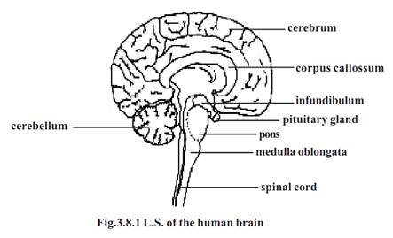
The human brain weighs about 1.3 Kg. It contains more than a billion neurons. Based on embryological development the brain can be divided as follows.
1-Prosencephalon (Fore brain) - It consists of the cerebrum and the diencephalon. The cerebrum is the largest part of the brain. It is divided into right and left hemispheres by a longitudinal fissure. However, at the base the two hemispheres are connected by a sheet of nerve fibres called the corpus callossum.
The outer surface of the cerebrum is called the cortex or grey mater. It is 2 to 4 mm thick. The inner content of the cerebrum is the white mater. The surface of the cerebrum has several folds called the gyri. They greatly increase the surface area of the cortex. The shallow grooves in between the gyri are called the sulci. A central sulcus runs in the lateral surface of the cerebrum from superior to inferior region.
Each cerebral hemisphere is divided into four lobes. They are the frontal at the front, the parietal towards the top of the head, the temporal on the side and the occipital at the rear.
The diencephalon contains the thalamus and hypothalamus. This region is found between the cerebrum and the brain stem.
The thalamus has a cluster of nuclei which act as the relays for particular sensory pathways. Just beneath the thalamus, the hypothalamus is present. It contains reflex centres linked to the autonomic system. A funnel shaped stalk called the infundibulum extends from its floor. It is connected to the neurohypophysis of the pituitary gland.
2-Mesencephalon (mid brain) - It is the smallest region of the brainstem. On its dorsal surface there are four rounded bodies called the carpora quadrigemina.
3-Rhombencephalon (hind brain) - The three main regions of the rhombencephalon are the medulla oblongata, the pons varoli and the cerebellum.
The cerebellum consists of two hemispheres. Its surface has many ridges called folia. The cerebellum consists of three parts. They are the small anterior flocconodular lobe, a narrow central vermis and two large lateral hemispheres.
The pons is just superior to the medulla oblongata. It contains ascending and descending nerve tracts.
The medulla oblongata is about 3 cm long. It is continuous with the spinal cord. It remains as a bridge between the brain and the spinal cord.
Brains stem - The medulla oblongata, pons and mid brain form the brain stem. It connects the spinal cord to the brain. Ten of the twelve cranial nerves enter or exit the brain through the brain stem.
Spinal cord - The spinal cord extends from the foramen magnum to the level of the second lumbar vertebra. It is considerably shorter than the vertebral column. There are two enlargements in the spinal cord. They are the cervical and lumbar enlargements. Below the lumbar enlargement the spinal cord tapers to form a cone like region called the conus medullaris. A connective tissue filament called the filum terminale extends inferiorly from conus medullaris to the coccyx. The conus medullaris and the nerves extending below resemble a horse’s tail. Hence it is called cauda equina.
A cross section of the spinal cord reveals a central grey portion and a peripheral white portion. The white matter consists of nerve tracts and the grey matter consists of neuron cell bodies and dendrites.
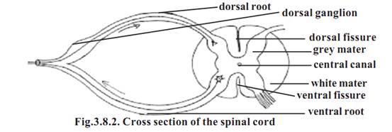
The dorsal and ventral sides have long fissures. There are 31 pairs of spinal nerves arising from the spinal cord. Each nerve has a dorsal root and a ventral root from the spinal cord. The dorsal roots have dorsal root ganglia.
Ventricles
The entire CNS remains as a hollow tube. The tube inside the adult brain forms ventricles.
Each cerebral hemisphere contains a large cavity called the lateral ventricle. It corresponds to the hypothetical first and second ventricles. The two lateral ventricles communicate with the third ventricle located in the center of the diencephalon. This connection is made through two interventricular foramina (foramen of Monro). The third ventricle in turn opens into the fourth ventricle found inside the medulla oblongata. This communication happens through a narrow canal called the cerebral aqueduct (aqueduct of sylvius). The fourth ventricle is continuous with the central canal of the spinal cord. The central canal extends nearly to the full length of the cord.
Cerebrospinal fluid (CSF)
This fluid fills the ventricles of the brain and the central canal of the spinal cord. About 80-90 % of CSF is produced by specialized cells called ependymal cells within the lateral ventricles. Remaining 10-12 % is produced by similar cells in the 3rd and 4th ventricles. These ependymal cells, their supportive tissue and the associated blood vessels together are called choroid plexuses. The plexuses are formed by invagination of the vascular piamater into the ventricles.
References
T. Sargunam Stephen, Biology (Zoology). First Edition – 2005, Government of Tamilnadu.
 الاكثر قراءة في علم التشريح
 اخر الاخبار
اخبار العتبة العباسية المقدسة

الآخبار الصحية


 قسم الشؤون الفكرية يصدر كتاباً يوثق تاريخ السدانة في العتبة العباسية المقدسة
 "المهمة".. إصدار قصصي يوثّق القصص الفائزة في مسابقة فتوى الدفاع المقدسة للقصة القصيرة
 (نوافذ).. إصدار أدبي يوثق القصص الفائزة في مسابقة الإمام العسكري (عليه السلام)


