


النبات

مواضيع عامة في علم النبات
الجذور - السيقان - الأوراق
النباتات الوعائية واللاوعائية
البذور (مغطاة البذور - عاريات البذور)
الطحالب
النباتات الطبية
الحيوان
مواضيع عامة في علم الحيوان
علم التشريح
التنوع الإحيائي
البايلوجيا الخلوية
الأحياء المجهرية
البكتيريا
الفطريات
الطفيليات
الفايروسات
علم الأمراض
الاورام
الامراض الوراثية
الامراض المناعية
الامراض المدارية
اضطرابات الدورة الدموية
مواضيع عامة في علم الامراض
الحشرات
التقانة الإحيائية
مواضيع عامة في التقانة الإحيائية
التقنية الحيوية المكروبية
التقنية الحيوية والميكروبات
الفعاليات الحيوية
وراثة الاحياء المجهرية
تصنيف الاحياء المجهرية
الاحياء المجهرية في الطبيعة
أيض الاجهاد
التقنية الحيوية والبيئة
التقنية الحيوية والطب
التقنية الحيوية والزراعة
التقنية الحيوية والصناعة
التقنية الحيوية والطاقة
البحار والطحالب الصغيرة
عزل البروتين
هندسة الجينات
التقنية الحياتية النانوية
مفاهيم التقنية الحيوية النانوية
التراكيب النانوية والمجاهر المستخدمة في رؤيتها
تصنيع وتخليق المواد النانوية
تطبيقات التقنية النانوية والحيوية النانوية
الرقائق والمتحسسات الحيوية
المصفوفات المجهرية وحاسوب الدنا
اللقاحات
البيئة والتلوث
علم الأجنة
اعضاء التكاثر وتشكل الاعراس
الاخصاب
التشطر
العصيبة وتشكل الجسيدات
تشكل اللواحق الجنينية
تكون المعيدة وظهور الطبقات الجنينية
مقدمة لعلم الاجنة
الأحياء الجزيئي
مواضيع عامة في الاحياء الجزيئي
علم وظائف الأعضاء
الغدد
مواضيع عامة في الغدد
الغدد الصم و هرموناتها
الجسم تحت السريري
الغدة النخامية
الغدة الكظرية
الغدة التناسلية
الغدة الدرقية والجار الدرقية
الغدة البنكرياسية
الغدة الصنوبرية
مواضيع عامة في علم وظائف الاعضاء
الخلية الحيوانية
الجهاز العصبي
أعضاء الحس
الجهاز العضلي
السوائل الجسمية
الجهاز الدوري والليمف
الجهاز التنفسي
الجهاز الهضمي
الجهاز البولي
المضادات الميكروبية
مواضيع عامة في المضادات الميكروبية
مضادات البكتيريا
مضادات الفطريات
مضادات الطفيليات
مضادات الفايروسات
علم الخلية
الوراثة
الأحياء العامة
المناعة
التحليلات المرضية
الكيمياء الحيوية
مواضيع متنوعة أخرى
الانزيمات

Face
 المؤلف:
Kelly M. Harrell and Ronald Dudek
المؤلف:
Kelly M. Harrell and Ronald Dudek
 المصدر:
Lippincott Illustrated Reviews: Anatomy
المصدر:
Lippincott Illustrated Reviews: Anatomy
 الجزء والصفحة:
 27-7-2021
 5348
5348


+
-
20
Face
The face, which spans between the ears and extends superior to inferior from the forehead to the chin, represents the anterior portion of the head. The general shape of the face is determined by the underlying bony scaffold of the viscerocranium and the overlying muscles, fascia, subcutaneous adipose, and skin. Cartilaginous extensions of the nose, eyelids, and external ears also provide unique features to each person's face.
A. Embryology
The pharyngeal apparatus gives rise to adult head and neck structures. For completeness, all head and neck adult derivatives are mentioned in this chapter with details on structures of the head.
1. Pharyngeal apparatus development: The pharyngeal apparatus contributes greatly to the formation of the head and neck region of the body (Figs. 1 and 2). The pharyngeal apparatus consists of the pharyngeal arches (five), pouches (four), grooves (four), and membranes (four). At week 4, the pharyngeal apparatus is first observed and gives the embryo its distinctive appearance. The particular fates of the elements of the pharyngeal apparatus are described below and shown in Table 1.
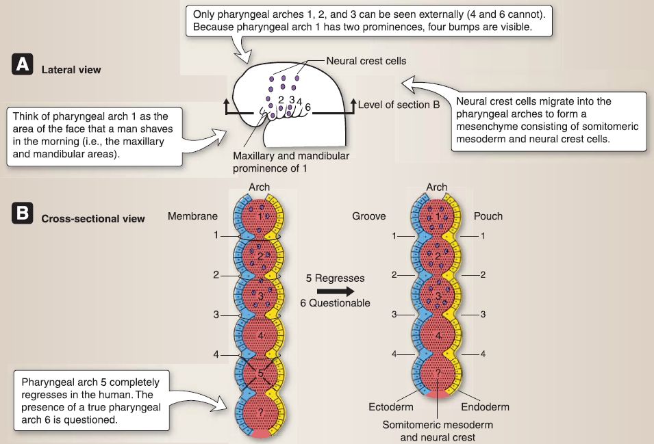
Figure 1: Pharyngeal apparatus.
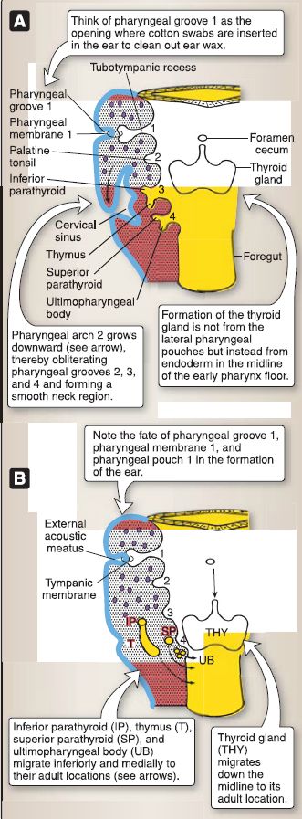
Figure 2: Pharyngeal pouch,groove,and membrane fate. A, 5 weeks. B, 7 weeks.
Table 1: Fate of Pharyngeal Apparatus
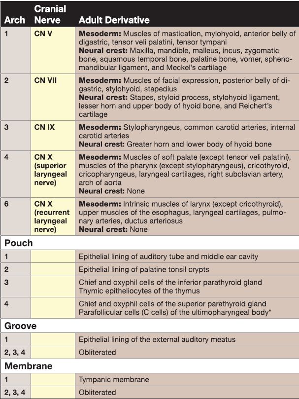
a. Pharyngeal arches: The pharyngeal arches (1, 2, 3, 4, and 6) contain somitomeric mesoderm and neural crest cells. The somitomeric mesoderm differentiates into various adult muscles and arteries (i.e., aortic arches 1-6) in the head and neck region. The neural crest cells differentiate into various adult bones and connective tissue in the head and neck region. In addition, each pharyngeal arch has a cranial nerve associated with it.
b. Pharyngeal pouches: The pharyngeal pouches (1, 2, 3, 4) are evaginations of the endoderm that lines the foregut.
[1] Pharyngeal pouch 1: This pouch elongates toward the surface and forms the tubotympanic recess. The endoderm lining the tubotympanic recess eventually forms the epithelial lining of the auditory tube and the middle ear cavity.
[2] Pharyngeal pouch 2: This pouch grows into the underlying mesenchyme and forms pitlike depressions that are lined by endoderm. This endoderm eventually forms the epithelial lining of the palatine tonsil crypts. The mesenchyme surrounding the crypts later becomes populated by lymphoid tissue forming the palatine tonsil.
[3] Pharyngeal pouch 3: This pouch expands and forms a solid dorsal portion related to the inferior parathyroid gland and a hollow ventral portion related to the thymus gland.
(a) Dorsal portion: The endoderm lining the solid dorsal portion progressively differentiates and eventually forms the chief cells and oxyphil cells of the inferior parathyroid gland. The bilateral primordia of the inferior parathyroid glands migrate inferiorly and medially where they come to lie on the dorsal surface of the lower part of the thyroid gland.
(b) Ventral portion: The endoderm lining the hollow ventral portion proliferates and forms solid cords of cells that grow into the underlying mesenchyme. The endoderm progressively differentiates and eventually forms the thymic epitheliocytes of the thymus gland. The mesenchyme surrounding the solid cords of cells later becomes populated by lymphoid tissue. The bilateral primordia of the thymus migrate inferiorly and medially where they fuse to form the bilobed thymus gland, which then descends into the superior mediastinum.
[4] Pharyngeal pouch 4: This pouch expands and forms a solid dorsal portion related to the superior parathyroid gland and an elongated ventral portion related to the ultimopharyngeal body.
(a) Dorsal portion: The endoderm lining the solid dorsal portion progressively differentiates and eventually forms the chief cells and oxyphil cells of the superior parathyroid gland. The bilateral primordia of the superior parathyroid glands migrate inferiorly and medially where they come to lie on the dorsal surface of the upper part
of the thyroid gland.
(b) Ventral portion: Very soon after the elongated ventral portion forms, it becomes populated by neural crest cells that form the ultimopharyngeal body. The bilateral ultimopharyngeal bodies detach from the pharyngeal wall and fuse with the dorsal surface of the thyroid gland. The neural crest cells within the ultimopharyngeal bodies
disperse throughout the thyroid gland and differentiate into the parafollicular cells (or C cells).
c. Pharyngeal grooves: The pharyngeal grooves (1, 2, 3, 4) are invaginations of the ectoderm that covers the surface of the embryo. They are located between each pharyngeal arch (see Fig. 2).
[1] Pharyngeal groove 1: This groove elongates toward the pharynx, and the ectoderm forms the epithelial lining of the external auditory meatus.
[2] Pharyngeal grooves 2, 3, and 4: These grooves are obliterated as pharyngeal arch 2 grows downward and fuses with the lower portion of the neck, thereby forming the smooth, contoured surface seen in the adult.
d. Pharyngeal membranes: The pharyngeal membranes (1, 2, 3, 4) are structures consisting of ectoderm, intervening mesoderm and neural crest, and endoderm. They are located between each pharyngeal arch. The first membrane is the only membrane that develops into an adult structure, the tympanic membrane. Membranes 2-4 become obliterated with the development of the neck and, therefore, are not represented as adult structures.
2. Face development: During weeks 4-10, the face and the palate form via five swellings: one frontonasal prominence, two maxillary prominences (from pharyngeal arch 1 ), and two mandibular prominences (from pharyngeal arch 1 ), as shown in Figure 3. The five swellings grow ventrally and medially due to the infiltration of neural crest cells and join each other in the midline of the future face. The five swellings surround the primitive oral cavity (stomodeum), which is separated from the gastrointestinal tract by the oropharyngeal membrane. Palate and nasal septum development will be detailed in "Oral" and "Nasal cavity" sections, respectively.
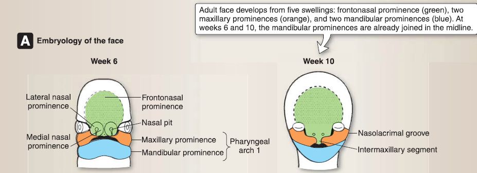
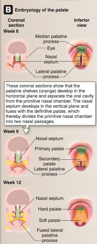
Figure 3: A, Face embryology at 6 and 10 weeks. B, Embryology of the palate at 6, 9, and 12 weeks.
a. Nasal placodes: During week 5, bilateral ectodermal thickenings called nasal placodes develop on the ventrolateral aspects of the frontonasal prominence. The nasal placodes invaginate into the underlying mesenchyme to form the nasal pits, thereby producing ridges of tissue that form the medial nasal prominences and lateral nasal prominences.
b. Nasolacrimal grooves: Deep grooves called the nasolacrimal grooves form between the maxillary prominences and the lateral nasal prominences and eventually form the nasolacrimal ducts and lacrimal sacs.
B. Musculature
Muscles of the face arise from bone and attach directly into fascia and skin of the face and neck (Fig. 4). Primarily, these muscles surround orifices-ears, nose, eyes, and mouth-and function as either dilators or sphincters at these openings. Secondarily, facial muscles move the skin of the face and neck in order to express a myriad of emotions. The main muscles of the face and their actions are described below.
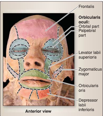
Figure 4: Face muscles.
1. Orbicularis oculi: This muscle is the sphincter of the eye. Its palpebral portion closes the eyelid gently, and its orbital portion closes the eyelid tightly.
2. Orbicularis oris: This muscle is the sphincter of the mouth. It closes and protrudes the lips.
3. Buccinator: The cheek muscle keeps the bolus of food between the teeth during mastication.
4. Zygomaticus major: The "smile muscle" pulls the angle of the mouth superiorly and laterally.
5. Other muscles: A number of smaller muscles around the eye, nose, ear, and mouth are devoted to actions, including lip elevation and depression, nostril dilation, and skin wrinkling on the bridge of the nose and chin. Although these muscles are not detailed here, their anatomical arrangement is partially shown in Figure 4 .
C. Innervation
Muscles of facial expression are derived from the second pharyngeal arch, so it makes sense that they receive motor innervation from the CN also associated with the second arch-facial nerve (CN VII), as shown in Figures 5. The face receives sensory innervation from the trigeminal nerve (CN V) (Fig. 6).
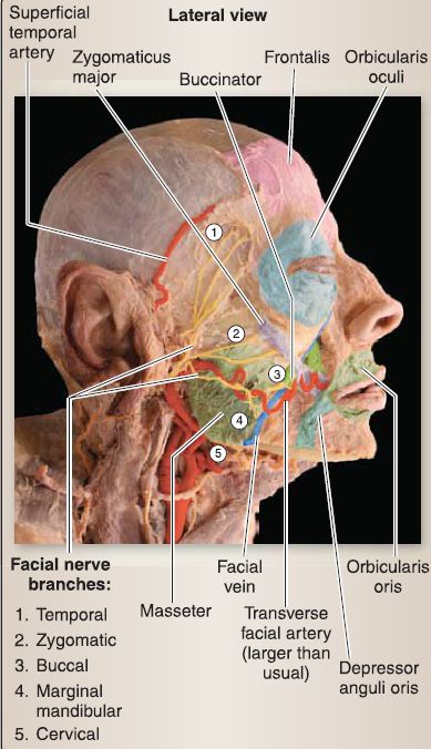
Figure 5: Face motor innervation. Facial nerve brances (CN VII).
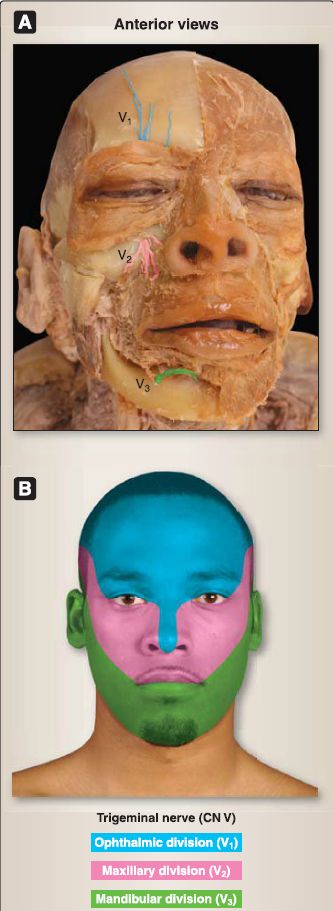
Figure 6: Face sensory innervation. Terminal branches of trigeminal nerve (CN V) divisions. A, Cadaveric specimen showing distal fibers. B, Trigeminal division distribution.
1. Facial nerve branches: The facial nerve provides branchiomotor (special visceral efferent [SVE]) innervation to all muscles of facial expression through regional terminal branches that reach the face after passing through the substance of the parotid gland. The five branches are temporal, zygomatic, buccal, marginal mandibular, and cervical.
2. Trigeminal nerve branches: Sensory (general somatic afferent [GSA]) innervation of the face is supplied by branches of the trigeminal nerve (CN V).
a. CN V1: Supratrochlear, supraorbital, infratrochlear, and external nasal nerves supply the skin over the upper eyelids, bridge of the nose, and forehead.
b. CN V2: lnfraorbital, zygomaticofacial, and zygomaticotemporal nerves supply the skin over the lateral nose, superior lip, inferior eyelids, cheekbone, and anterior temporal fossa.
c. CN V3: Mental, buccal, and auriculotemporal nerves supply the skin over the inferior lip, chin, flesh of the cheek, and temporal fossa and parotid regions.
D. Vasculature
The face is primarily supplied by direct or indirect branches of the external carotid artery. Venous tributaries of the face typically drain into the jugular venous system.
1. Arterial supply: Superficial structures of the face are primarily supplied by the facial artery, a direct branch of the external carotid artery (Fig. 7). From its origin in the carotid triangle of the neck, the facial artery follows a tortuous path, deep to the superficial part of the submandibular gland and crosses the inferior border of the mandible, anterior to the masseter muscle. At the angle of the mouth, it gives off inferior and superior labial arteries, to supply the structures of the lower and upper lips, respectively. The facial artery continues toward the nose, where it is renamed the angular artery. Additional arteries that supply the face include branches of the superficial temporal artery.
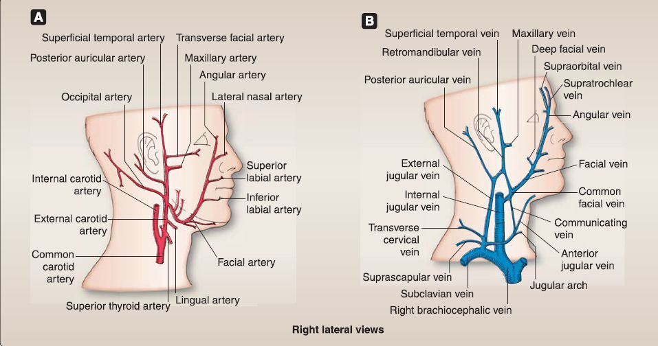 Figure 7: Blood supply to face A, Arterial supply. B, Venous supply.
2. Venous supply: Tributaries of the facial vein drain the majority of the face, including the angular, superior and inferior labial, and deep facial veins (see Fig. 7). The facial vein unites with the anterior branch of the retromandibular vein to form the common facial vein, which drains directly into the internal jugular vein. The posterior branch of the retromandibular vein joins the posterior auricular vein to form the external jugular vein.
3. Lymphatic supply: Lymph nodes of the face are only found in the parotid and buccal regions (Fig. 8). The majority of lymph is drained, along with lymph from the scalp, through the superficial ring of lymph nodes at the intersection of the head and neck (including parotid, occipital, mastoid, submental, and submandibular nodes). Once drained through the superficial ring, lymph travels through the deep cervical nodes adjacent to the internal jugular vein to the jugular lymphatic duct. Lymph eventually drains into the thoracic duct on the left of the subclavian/internal jugular vein junction on the right.
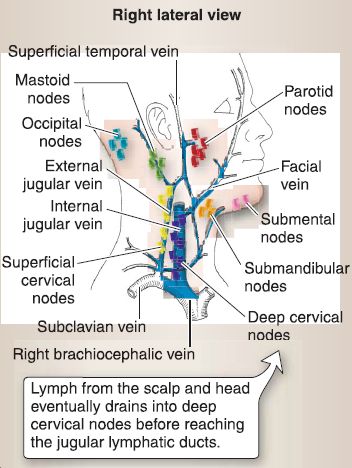
Figure 8: Head lymphatics. Figure showing primary lymph nodes of head.
 الاكثر قراءة في علم التشريح
 اخر الاخبار
اخبار العتبة العباسية المقدسة

الآخبار الصحية


 قسم الشؤون الفكرية يصدر كتاباً يوثق تاريخ السدانة في العتبة العباسية المقدسة
 "المهمة".. إصدار قصصي يوثّق القصص الفائزة في مسابقة فتوى الدفاع المقدسة للقصة القصيرة
 (نوافذ).. إصدار أدبي يوثق القصص الفائزة في مسابقة الإمام العسكري (عليه السلام)


