Embryology of Upper Limb
At the end of week 4, the upper limb buds form pronounced protrusions from the lateral body wall at the C5-T2 level (Fig. 1). In general, the upper limb bud consists of a core of mesoderm that is covered by ectoderm. Mesoderm from the lateral plate (lateral plate mesoderm) migrates into the upper limb bud and condenses in the center of the limb bud to eventually form the skeletal component (i.e., bone, ligaments, tendons, and dermis) of the upper limb. Mesoderm from the somites (somitomeric mesoderm) migrates into the upper limb bud and condenses into a posterior extensor condensation and an anterior flexor condensation to eventually form the muscular component of the upper limb.
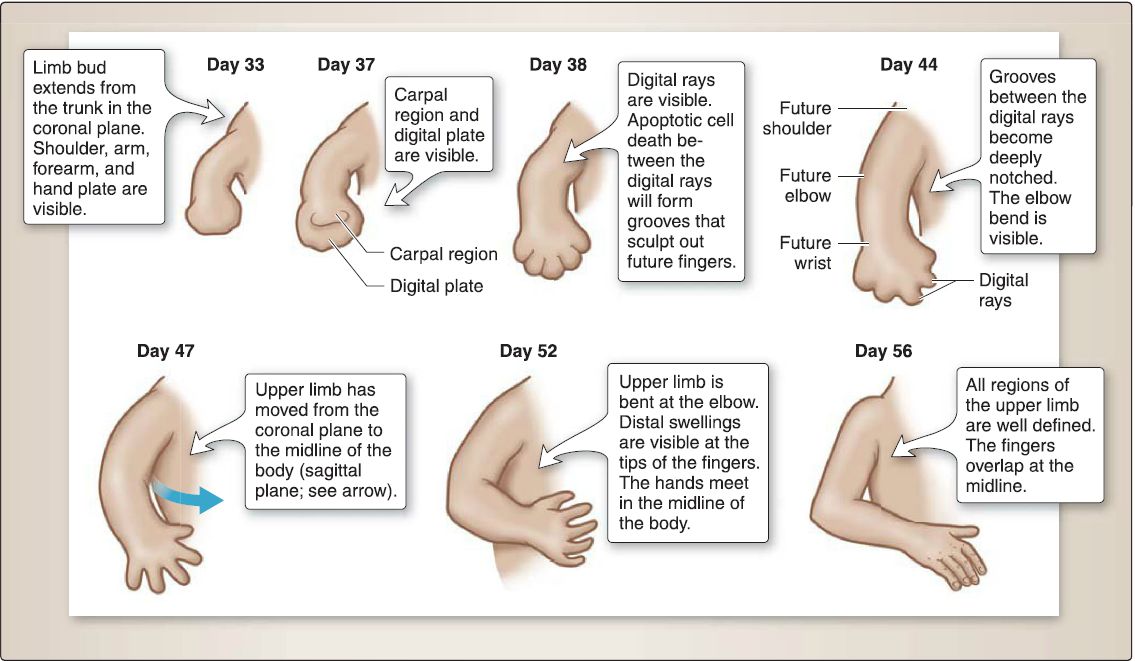 Figure 1: Embryologic development of the upper limb.
A. Special development zones
The upper limb bud displays two specialized areas called the zone of polarizing activity (ZPA) and the apical ectodermal ridge (AER) that play an important role in the development of the upper limb. The ZPA consists of mesodermal cells located just beneath the AER along the caudal margin of the upper limb bud. The AER is a thickened ridge of ectoderm at the tip of the upper limb bud.
B. Development axes
The upper limb bud develops with respect to three axes. The proximaldistal axis runs from the shoulder to the fingers. The cranial-caudal axis runs from the thumb to the little finger. The posterior-anterior axis runs from the back of the hand to the palm of the hand.
C. Bone formation
The lateral plate mesoderm migrates into the upper limb bud and condenses in the center of the upper limb bud to eventually form the skeletal component of the upper limb. The lateral plate mesoderm forms the scapula, clavicle, humerus, radius, ulna, carpals, metacarpals, and phalanges. All the bones of the upper limb undergo endochondral ossification. However, the clavicle undergoes both membranous and endochondral ossification.
D. Muscle formation
The upper limb bud site lies opposite somites C4-C8, T1, and T2. During week 5, mesoderm from these somites (myotomes) migrates into the limb bud and forms a posterior extensor condensation of mesoderm and an anterior flexor condensation of mesoderm. The mesoderm of these condensations differentiates into myoblasts. Then, the condensations split into anatomically recognizable muscles of the upper limb, although little is known about this process.
1. Posterior extensor condensation of mesoderm: In general, this gives rise to the extensor and supinator musculature (Table 1).
Table 1: Posterior and Anterior Mesoderm Condensations Into Muscle
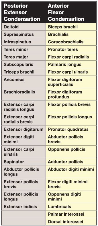
2. Anterior flexor condensation of mesoderm: In general, this gives rise to the flexor and pronator musculature (see Table 1 ).
E. Brachial plexus formation
The axons within anterior primary rami from C5-C8 and T1 arrive at the base of the upper limb bud and mix in a specific pattern to form the upper trunk, middle trunk, and lower trunk of the brachial plexus.
1. Axon migration and directionality: At this point, axon migration pauses at the base of the upper limb bud and then subsequently resumes so that axons are directed to either the posterior extensor condensation or the anterior flexor condensation. The direction that the axons take is controlled by the homeobox gene Lim1 and its downstream target ephrin type A receptor 4 (EPHA4), allowing axons to enter the posterior extensor condensation but avoiding the anterior flexor condensation.
2. Cord formation: The posterior divisions of the trunks grow into the posterior extensor condensation of mesoderm and join to form the posterior cord. The anterior divisions of the trunks grow into the anterior flexor condensation of mesoderm and join to form the medial cord and lateral cord.
3. Terminal branch formation: With further development of the limb musculature, the posterior cord branches into the axillary nerve (C5, C6) and radial nerve (C5-C8, T1), thereby innervating all the muscles that form from the posterior extensor condensation of mesoderm. With further development of the limb musculature, the medial cord and lateral cord branch into terminal nerves including the musculocutaneous nerve (C5-C7), ulnar nerve (C8, T1), and median nerve (C5-C8, T1), thereby innervating all the muscles that form from the anterior flexor condensation of mesoderm.
F. Upper limb vasculature formation
The upper limb vasculature formation involves the fates of the subclavian artery and the axis artery.
1. Fate of the subclavian artery: The subclavian artery enters the upper limb bud as the axis artery, which ends in a terminal plexus near the tip of the upper limb bud. The terminal plexus participates in the formation of the deep palmar arch and the superficial palmar arch.
2. Fate of the axis artery: The axis artery initially sprouts the posterior interosseous artery and the median artery. The median artery is typically regresses in the adult. The axis artery later sprouts the radial artery and ulnar artery. The axis artery persists in the adult as the axillary artery, brachia! artery, anterior interosseous artery, and deep palmar arch.
G. Rotation and dermatome pattern
At week 4, the upper limb buds appear as small bulges oriented in the coronal plane (Fig. 2). In week 6, the upper limb buds undergo a horizontal movement, so that they are now oriented in the sagittal plane. During weeks 6-8, the upper limbs undergo a 90° lateral rotation, such that the elbow points posteriorly, the extensor compartment lies posterior, and the flexor compartment lies anterior. The 90° lateral rotation of the upper limb bud alters the originally straight segmental pattern of innervation in the embryo; that is, the pattern of innervation becomes "twisted in a spiral" in the adult (Fig. 3).
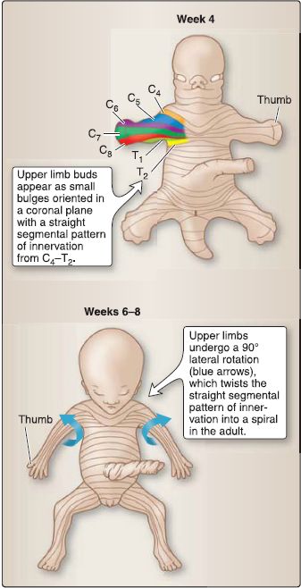
Figure 2: Rotation of the upper limb.
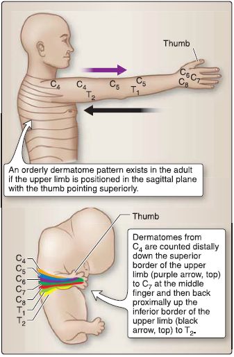
Figure 3: Dermatome pattern of the upper limb.


