Musculature
The muscles of the lower limb are organized into specific compartments or regions: gluteal region; anterior, medial, and posterior thigh; and anterior, lateral, and posterior leg. The muscles in these compartments commonly share similar neurovascular supply and overall function(s), with a few exceptions.
A. Gluteal region
The gluteal region is associated with the pelvis and hip joint posteriorly. Superficially, the gluteal region contains great amounts of subcutaneous adipose tissue overlying a thick deep fascia that encompasses the underlying musculature. The deep fascia of the gluteal region is continuous with the fascia lata but separates the gluteal region from the posterior thigh. Musculature of the gluteal region is divided into two groups-superficial and deep.
1. Superficial group: The superficial group comprises three gluteal muscles-gluteus maximus, medius, and minimus-and tensor fascia latae.
a. Gluteus maximus: This muscle is rhomboid-shaped and covers all other gluteal region muscles except for the superior fibers of gluteus medius along the posterior ilium (Fig. 1). Gluteus maxim us is a powerful extensor of the femur and also assists in lateral rotation and adduction (inferior fibers) of the femur. It is innervated by the inferior gluteal nerve and supplied by both the superior and inferior gluteal vessels.
b. Gluteus medius and minimus: These are fan-shaped muscles that abduct and medially rotate the femur (Fig. 2). Additionally, and very importantly, gluteus medius and minim us function together to maintain a level pelvis during the gait cycle. Both muscles are innervated and supplied by the superior gluteal nerve and vessels, respectively.
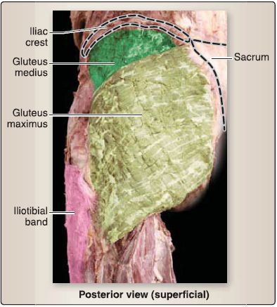
Figure 1: Superficial muscles of the gluteal region.
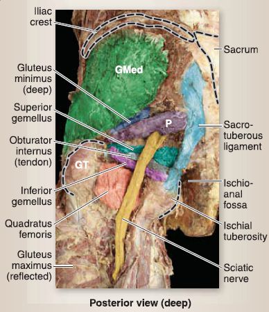
Figure 2 : Deep muscles of the gluteal region. GMed = gluteus medius, GT = greater
trochanter, P = piriformis.
c. Tensor fascia latae: While included in this group, the tensor fascia latae is somewhat of a transitional muscle, as it is positioned between the gluteal region and the anterior thigh. It functions to abduct, flex, and medially rotate the femur and is innervated by the superior gluteal nerve. Along with gluteus maximus, tensor fascia latae attaches distally via the iliotibial band, and therefore, these muscles actively assist in stabilizing
the knee joint as well.
2. Deep musculature: The deep group comprises a collection of small muscles that laterally rotate the femur and stabilize the hip joint (see Fig. 2). These muscles include piriformis, superior gemellus, obturator internus, inferior gemellus, and quadratus femoris. Each of these muscles is innervated by nerves of the same name that arise from the lumbosacral plexus (i.e., nerve to piriformis; nerve to superior gemellus and obturator internus; nerve to inferior gemellus and quadratus femoris). These five muscles collectively function to laterally rotate the femur and stabilize the head of the femur in the acetabulum.
a. Piriformis: The pear-shaped piriformis is the most superior of the group and serves as a landmark structure in the gluteal region. Superior and inferior gluteal neurovascular bundles exit the pelvis superior and inferior to the piriformis, respectively. The sciatic nerve and posterior femoral cutaneous nerve also exit the pelvis inferior to the piriformis.
b. Superior and inferior gemelli and obturator internus: These muscles flank the tendon of the obturator internus as all three muscles insert on the greater trochanter.
c. Quadratus femoris: This is the most inferior of the deep muscles, and it inserts on the intertrochanteric crest of the femur.
3. Cutaneous innervation: Cutaneous innervation of the gluteal region is mediated by cluneal nerves. From the iliac crest to the gluteal crease, dermatomes L3-L5 and S1-S5 are represented in the gluteal region (Fig. 3).
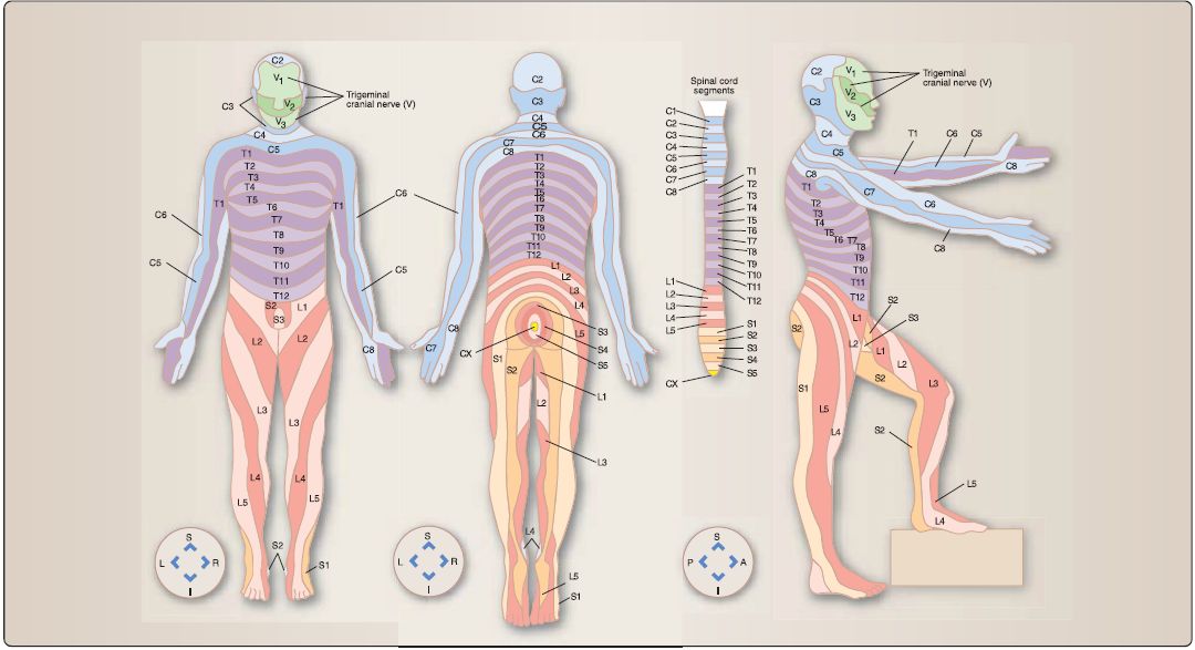 Figure 3 : Dermatome maps. A = anterior, I = inferior, L = left, P = posterior, R = right, S = superior.
B. Thigh
The thigh region spans between the hip and knee joints. The femur is the primary bone of the thigh, although muscles of this region also attach to the pelvis, tibia, and fibula and act at both the hip and knee joints. The fascia lata encases the thigh structures and gives rise to intermuscular septa, which divide this region into three main compartments-anterior, medial, and posterior. Each compartment has a main nerve and blood supply as well as muscles with common functions.
1. Anterior thigh: As shown in Figure 4, muscles of the anterior thigh include quadriceps femoris, iliopsoas, sartorius, and pectineus. In general, this group of muscles receives innervation from the femoral nerve (L2-L4) and functions to flex the hip and extend the knee. The femoral artery and its branches vascularize the anterior compartment.
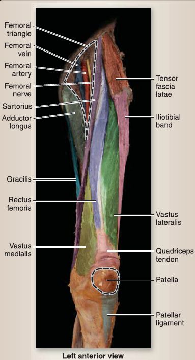
Figure 4: Anterior thigh.
a. Quadriceps femoris: This group of four muscles-rectus femoris, vastus lateralis, vastus medialis, and vastus intermedius-function together to extend the knee. All four
muscles insert through a common quadriceps tendon on the tibial tuberosity.
b. lliopsoas: The rectus femoris also crosses the hip joint and assists the iliopsoas with hip flexion. The iliopsoas is the primary flexor of the hip. In the anterior thigh, the iliopsoas represents the inferior portions of two muscles that originate in the posterior
body wall and greater pelvis-psoas major and iliacus, respectively. As a result of its proximal attachments, the iliopsoas is also active during standing to assist in maintenance of posture and lumbar curvature.
c. Sartorius (tailor's muscle): This passes obliquely across the anterior thigh from lateral to medial, crossing both the hip and knee joints. It flexes, abducts, and externally rotates the hip and flexes the knee.
d. Pectineus: This is a transitional muscle, between the anterior and medial thigh compartments. It adducts, flexes, and medially rotates the hip.
e. Femoral triangle: The anterior thigh contains important neurovascular structures that travel from the pelvis to the lower limb. These structures are arranged within a proximal space called the femoral triangle (Fig. 5). From lateral to medial, the triangle contains the femoral nerve, femoral artery, femoral vein, and lymphatics. The femoral artery, vein, and lymphatics are all contained in a funnel-shaped fascial covering called the femoral sheath, which is divided into lateral, intermediate, and medial compartments. The lateral compartment contains the femoral artery, intermediate the femoral vein, and medial compartment represents the femoral canal. The femoral canal contains varying amounts of loose connective tissue, fat, and lymphatics and communicates with the abdominal cavity. The boundaries of the femoral triangle are:
• Lateral: sartorius muscle
• Medial: adductor longus muscle
• Superior: inguinal ligament
• Floor: iliopsoas and pectineus muscles
• Roof: fascia lata
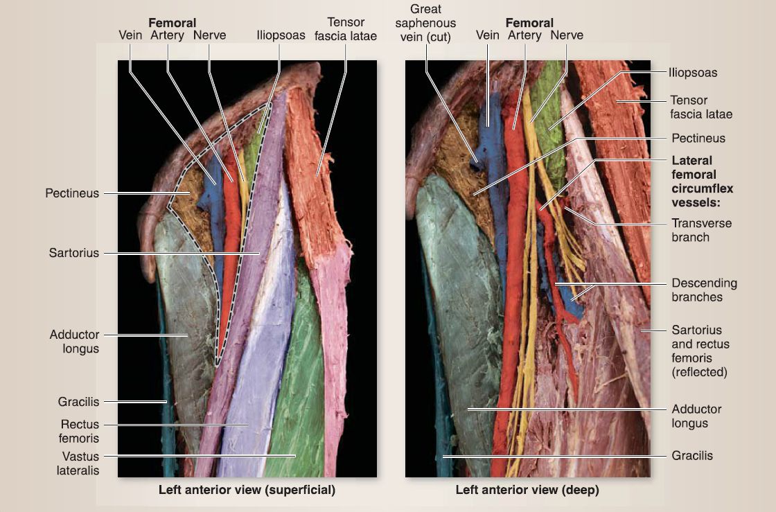 Figure 5: Femoral triangle.
f. Adductor canal (Hunter canal): This begins at the apex of the femoral triangle, at which point the femoral artery and vein, saphenous nerve, and nerve to the vastus medialis travel deep to the sartorius muscle. The canal provides an intermuscular passageway for the femoral artery and vein as they travel toward the adductor hiatus, a musculotendinous opening in the adductor magnus muscle. The boundaries of the adductor canal are:
• Posteromedial: adductor longus and adductor magnus
• Anterior: sartorius
• Lateral: vastus medialis
g. Cutaneous innervation: Cutaneous innervation of the anterior thigh is mediated by branches of the femoral (anterior branches) nerve, genitofemoral nerve, and lateral cutaneous nerve of the thigh. From the inguinal crease (hip crease) to the knee, dermatomes L1-L5 course lateral to medial across the anterior thigh .
2. Medial thigh: As shown in Figure 6, muscles of the medial thigh include adductor longus, adductor brevis, adductor magnus, gracilis, and obturator externus. Collectively, these muscles are innervated by the obturator nerve (L2-L4) and receive blood supply from the obturator artery-a branch of the internal iliac artery. In general, this group of muscles adducts the hip and plays a minor role in hip flexion.
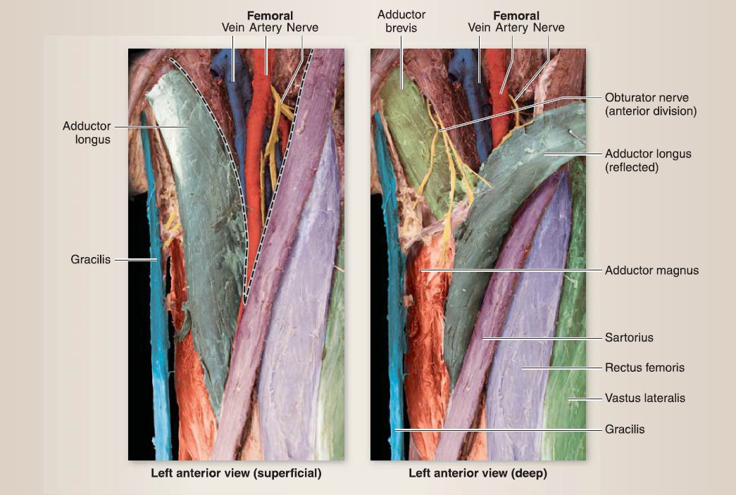 Figure 6: Medial thigh.
a. Adductors: The adductor longus is superficial to adductor brevis, and both arise from the pubis to insert along the medial femur. Adductor magnus is the largest muscle in the medial thigh and is made up of two parts-a horizontal adductor part, which is innervated by the obturator nerve, and a vertical hamstring part, which is innervated by the tibial division of the sciatic nerve. The adductor portion of adductor magnus arises from the pubis and adducts the hip, whereas the hamstring portion arises from the ischial tuberosity and extends the hip . The hamstring portion inserts medially on the adductor tubercle, which creates an opening in the tendon, previously described as the adductor hiatus.
b. Gracilis: This long, slender muscle crosses both the hip and knee joints and is most medial of the group. In addition to hip adduction, gracilis flexes the knee and medially rotates the hip.
c. Obturator externus: Located deep and superiorly in the medial compartment, obturator externus is not a hip adductor, but rather acts to externally rotate the hip, much like its counterpart obturator internus (V.A).
d. Cutaneous innervation: Cutaneous innervation of the medial thigh is mediated by branches of the obturator, femoral (anterior branches), and ilioinguinal nerves. From the inguinal crease to the knee dermatomes, L1-L3 course lateral to medial to the medial thigh .
3. Posterior thigh: As shown in Figures 7 and 8, muscles of the posterior thigh include semitendinosus, semimembranosus, and biceps femoris (short and long heads). Collectively, these muscles are innervated by divisions of the sciatic nerve (tibial or common fibular) and receive blood supply from the perforating branches of the profunda femoris artery. In general, this group of muscles extends the hip and flexes the knee.
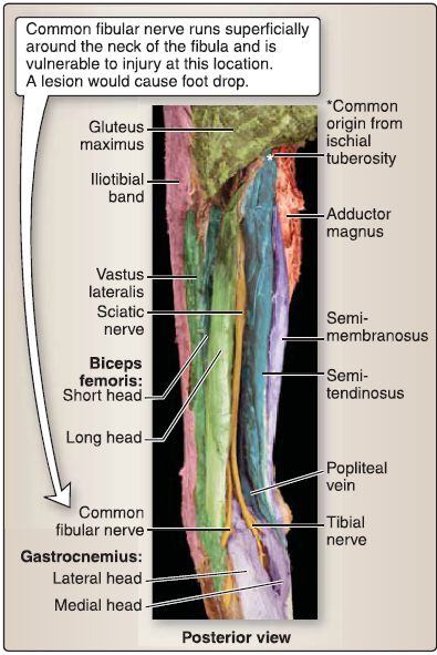
Figure 7: Posterior thigh.
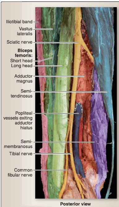
Figure 8:Posterior thigh and adductor hiatus {dotted line).
a. Semitendinosus, semimembranosus, and biceps femoris: Semitendinosus, semimembranosus, and biceps femoris (long head) muscles originate from the ischial tuberosity, cross the hip and knee joints, and insert on either the tibia or the fibula. They extend the hip and flex the knee and are innervated by the tibial division (L4,L5, S1-S3) of the sciatic nerve. Biceps femoris (short head) is the exception, as it arises directly from the shaft of the femur and only crosses the knee joint to assist in knee flexion. It is innervated by the common fibular (L4-L5, S1-S2) division of the sciatic nerve. Semitendinosus and semimembranosus are located medially in the posterior thigh, whereas biceps femoris is located laterally. The hamstring portion of adductor magnus lies anterior to semitendinosus and semimembranosus and also assists in hip extension, but not knee flexion, as it does not cross the knee joint. b. Cutaneous innervation: Cutaneous innervation of the posterior thigh is mediated by branches of the cluneal nerves and posterior and lateral cutaneous nerves of the thigh. From the gluteal crease to the knee, dermatomes L1-L2, L5, and S1-S2 course inferiorly along the posterior thigh .
C. Leg
The leg represents the portion of the lower limb between the knee and ankle. The tibia and fibula make up the main bones of the leg and are positioned medial and lateral, respectively. The crural fascia encases the leg structures and gives rise to intermuscular septa, which divide this region into three main compartments-anterior, lateral, and posterior. Each compartment has a main nerve and blood supply, as well as muscles with common functions.
1. Popliteal fossa: The popliteal fossa is a diamond-shaped space located on the distal posterior thigh and proximal posterior leg that frames the posterior knee joint and surrounding structures (Fig. 9). The popliteal fossa is bound superiorly by the hamstring muscles and inferiorly by the medial and lateral heads of gastrocnemius.The floor is made up of the posterior surface of the femur, posterior knee capsule, and popliteus muscle fascia. The roof is made up of skin and underlying deep fascia (popliteal fascia).
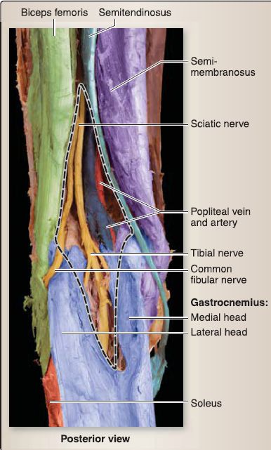
Figure 9: Popliteal fossa (dotted line).
a. Contents: The popliteal fossa is an important space, as it contains all of the major neurovascular structures traveling to/from the leg and foot. The contents are:
• Popliteal artery and vein and respective branches/tributaries
• Tibial and common fibular nerves
• Sural nerve branches
• Lymph vessels and nodes (superficial and deep popliteal)
• Fat
• Small saphenous vein (where it drains into the popliteal vein)
b. Nerves: The tibial nerve will continue inferiorly to innervate structures in the posterior compartment of the leg and plantar surface of the foot. The common fibular nerve will travel laterally to the level of the fibular head and split into deep and superficial fibular nerves to innervate structures in the anterior and lateral leg compartments, respectively.
2. Anterior leg: As shown in Figure 10, muscles of the anterior leg include tibialis anterior, extensor digitorum, extensor hallucis longus, and fibularis tertius. Muscles of the anterior leg span between the lateral tibial surface and the medial fibular surface, anterior to the interosseous membrane. Collectively, these muscles are innervated by the deep fibular nerve and receive blood supply from the anterior tibial artery. In general, this group of muscles dorsiflexes the ankle and extends the toes.
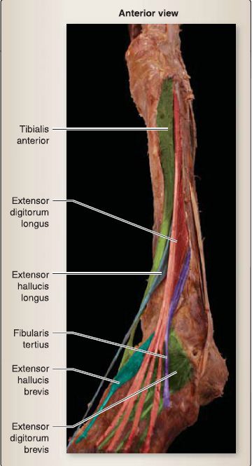
Figure 10: Anterior leg.
a. Tibialis anterior: Tibialis anterior is the primary ankle dorsiflexor and constitutes most of the muscle bulk in the anterior compartment.
b. Extensors: Extensor hallucis longus and extensor digitorum longus are positioned lateral to tibialis anterior and extend the hallux (first digit) and digits (second-fifth digits), respectively, as well as assist in ankle dorsiflexion.
c. Fibularis tertius: Although sometimes absent, when present, this muscle is adjacent to extensor digitorum and functions to dorsiflex the ankle and evert the foot.
d. Fascia-extensor retinacula: At the level of the distal anterior leg and ankle, extensor retinacula-superior and inferiortack down the tendons of the anterior leg muscles to prevent bow-stringing during contraction and joint movement. These retinacula are thickenings of the crural fascia that surrounds the leg musculature.
e. Cutaneous innervation: Cutaneous innervation of the anterior leg is mediated mainly by the saphenous nerve (femoral nerve branch) and partially by the lateral sural cutaneous nerve. From the knee to the ankle, dermatomes Lcl5 are represented in the anterior leg .
3. Lateral leg
Muscles of the lateral leg include fibularis longus and fibularis brevis and originate from the fibula (Fig. 11). Collectively, these muscles are innervated by the superficial fibular nerve and receive blood supply from the fibular artery. In general, this group of muscles everts the foot and weakly plantarflexes the ankle.
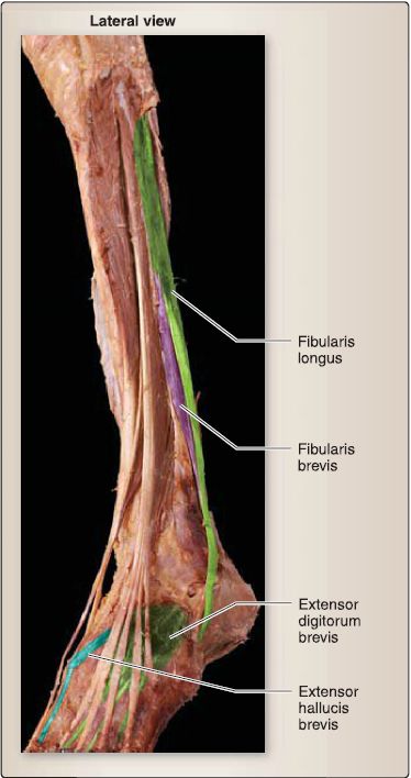
Figure 11: Lateral leg.
a. Fibularis longus and brevis: Fibularis longus sends its tendon across the plantar surface of the foot to insert medially. This attachment represents an active support of the transverse arch of the foot. Fibularis brevis inserts laterally on the base of the fifth metatarsal.
b. Fascia-fibular retinacula: At the level of the distal lateral leg and ankle, fibular retinacula-superior and inferior-tack down the tendons of the lateral leg muscles to prevent bow-stringing during contraction and joint movement. These retinacula are thickenings of the crural fascia that surrounds the leg musculature.
c. Cutaneous innervation: Cutaneous innervation of the lateral leg is mediated by the lateral sural cutaneous nerve and superficial fibular nerve. The L5 dermatome is represented along the lateral leg .
4. Posterior leg: Muscles of the posterior leg are arranged into superficial and deep groups and divided by the transverse intermuscular septum. Collectively, these muscles are innervated by the tibial nerve and receive blood supply from the posterior tibial and fibular arteries. In general, this group of muscles plantarflexes the ankle and flexes the toes.
a. Superficial muscles: Gastrocnemius, soleus, and plantaris share a common tendon-calcaneal ("Achilles") tendon that inserts on the calcaneus (Fig. 12). Muscles of the superficial posterior compartment represent the bulk of the "calf" muscle.

Figure 12: Posterior view (superficial) Superficial posterior leg.
[1] Gastrocnemius: The two-headed gastrocnemius is the most superficial and crosses both the knee and ankle. In addition to plantarflexing the ankle, the gastrocnemius flexes the knee.
[2] Soleus: Deep to the gastrocnemius is the fish-shaped soleus muscle, which only crosses the ankle joint and is, therefore, a powerful plantarflexor.
[3] Plantaris: When present, this muscle has a small muscle belly and long thin tendon, which limits its contribution to function.
b. Deep muscles: Popliteus, tibialis posterior, flexor digitorum, and flexor hallucis long us are the muscles of the deep posterior compartment and contribute to plantarflexion of the ankle as well as flexion of the toes (Fig. 13). From medial to lateral, flexor digitorum longus, tibialis posterior, and flexor hallucis longus arise from the posterior surface of the tibia, interosseous membrane, and fibula.
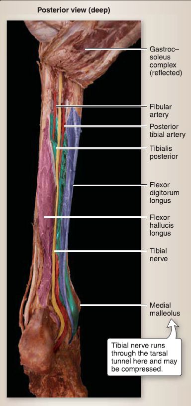
Figure 13: Deep posterior leg.
[1] Tibialis posterior: Similar to the fibularis longus, the tibialis posterior provides active support for the medial longitudinal arch of the foot during weight bearing.
[2] Flexor hallucis longus: This muscle is a powerful flexor of the hallux, which enables adequate "push off" during the gait cycle (preswing phase).
[3] Flexor digitorum longus: This muscle flexes digits two through five and assists in plantarflexion.
[4] Popliteus: The popliteus muscle lies in the proximal deep posterior leg and plays an important part in unlocking a fully extended knee joint. In an open-chain position (unfixed foot) popliteus internally rotates the tibia on the femur, while in a close-chain position (foot fixed as in during stance phase in gait cycle), the popliteus externally rotates the femur. Both of these actions unlock a fully extended knee.
c. Tarsal tunnel: Just prior to traveling posterior to the medial malleolus, the tendons of tibialis posterior and flexor digitorum longus cross (Fig. 14). Thus, the order of structures from anterior to posterior) traveling into the plantar foot is:
• Tibialis posterior
• Flexor digitorum long us
• Posterior tibial artery/veins and tibial nerve
• Flexor hallucis longus
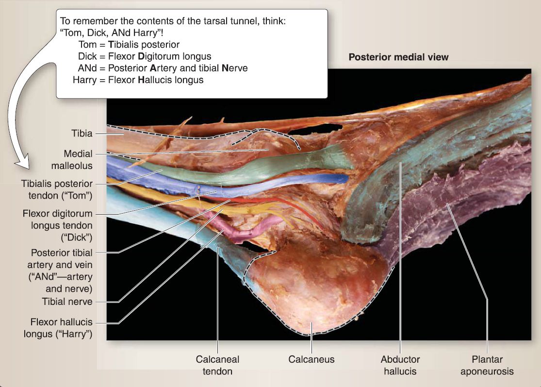 Figure 14 :Contents of the tarsal tunnel. Dotted line shows bony features.
d. Fascia-flexor retinaculum: At this level, the posterior tibial artery and tibial nerve divide into MP and LP arteries and nerves, respectively. A flexor retinaculum maintains the position of these structures posterior to the medial malleolus.
e. Cutaneous innervation: Cutaneous innervation of the posterior leg is mediated by the saphenous nerve (femoral nerve branch) and medial and lateral sural cutaneous nerves. From the knee to the ankle, dermatomes L3 and S1-S2 are represented in the posterior leg .
D. Ankle and foot
The ankle represents the articulation between the distal tibia and fibula, with the talus as the leg transitions into the foot. The foot is made up of 7 tarsal bones, 5 metatarsal bones, and 14 phalanges (proximal, middle, distal) and serves as a weight-bearing structure, important for standing and gait . The foot has three parts-hindfoot (talus and calcaneus), midfoot (navicular, cuboid, and cuneiforms), and forefoot (metatarsals and phalanges)-which are functionally important when discussing load bearing and movement during the gait cycle. When describing structures of the foot, it is further divided into dorsum and plantar regions.
1. Dorsum of foot: The dorsum of the foot contains two musclesextensor hallucis brevis and extensor digitorum brevis (Fig. 15). These muscles collectively extend the digits and arise from the calcaneus to insert onto the hallux and digits two through five, respectively. These muscles are innervated by the deep fibular nerve.
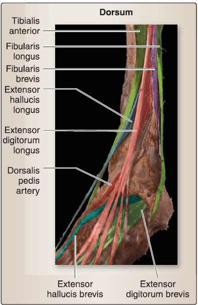
Figure 15: Dorsum of foot.
a. Vasculature: The main arterial supply to the dorsum of the foot is dorsalis pedis artery, which is the continuation of the anterior tibial artery in the leg. The dorsalis pedis artery travels distally, gives off tarsal branches, and terminates into the arcuate and deep plantar arteries. The dorsal venous network drains the dorsum of the foot.
b. Cutaneous innervation: Cutaneous innervation is mediated primarily by the superficial fibular nerve, while the deep fibular nerve supplies the skin between the first and second digits. Dermatomes L5-L4 and S1 are represented on the dorsum of the foot .
Plantar foot: The plantar surface of the foot houses most of the intrinsic foot muscles (Fig. 16). These muscles function more as a collective unit to constantly modify movement and stability during ambulation. Innervation is generally provided by the LP and MP nerves . The plantar foot is further divided into four muscular layers, arranged superficial to deep.
a. First layer: These muscles include abductor hallucis (MP), flexor digitorum brevis (MP), and abductor digiti minimi (LP). b. Second layer: These muscles include quadratus plantae (LP), lumbricals (MP/LP), and tendons of flexor hallucis longus and flexor digitorum longus.
c. Third layer: These muscles include flexor hallucis brevis (MP), adductor hallucis (LP), and flexor digiti minimi (LP).
d. Fourth layer: These muscles include plantar interossei (LP), dorsal interossei (LP), and tendons of fibularis longus and tibialis posterior.
e. Plantar aponeurosis: The protective deep fascia of the foot covers the plantar surface and thickens centrally into the plantar aponeurosis. The plantar aponeurosis extends from the calcaneus to the metatarsal heads and sends intermuscular septa superiorly to further compartmentalize the plantar surface of the foot.
f. Vasculature: Plantar blood supply is achieved through MP and LP arteries, which are the main terminal branches of the posterior tibial artery.
g. Cutaneous innervation: Cutaneous innervation is mediated primarily by the MP and LP nerves. The heel receives cutaneous innervation by way of the medial calcaneal branch of the tibial nerve. Dermatomes L5-L4 and S1-S2 are represented on the plantar surface of the foot .
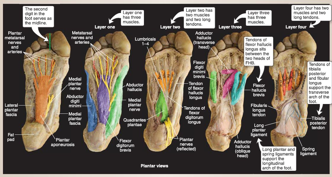 Figure 16: Layers of the foot. D = dorsal; P = plantar.


