


النبات

مواضيع عامة في علم النبات
الجذور - السيقان - الأوراق
النباتات الوعائية واللاوعائية
البذور (مغطاة البذور - عاريات البذور)
الطحالب
النباتات الطبية
الحيوان
مواضيع عامة في علم الحيوان
علم التشريح
التنوع الإحيائي
البايلوجيا الخلوية
الأحياء المجهرية
البكتيريا
الفطريات
الطفيليات
الفايروسات
علم الأمراض
الاورام
الامراض الوراثية
الامراض المناعية
الامراض المدارية
اضطرابات الدورة الدموية
مواضيع عامة في علم الامراض
الحشرات
التقانة الإحيائية
مواضيع عامة في التقانة الإحيائية
التقنية الحيوية المكروبية
التقنية الحيوية والميكروبات
الفعاليات الحيوية
وراثة الاحياء المجهرية
تصنيف الاحياء المجهرية
الاحياء المجهرية في الطبيعة
أيض الاجهاد
التقنية الحيوية والبيئة
التقنية الحيوية والطب
التقنية الحيوية والزراعة
التقنية الحيوية والصناعة
التقنية الحيوية والطاقة
البحار والطحالب الصغيرة
عزل البروتين
هندسة الجينات
التقنية الحياتية النانوية
مفاهيم التقنية الحيوية النانوية
التراكيب النانوية والمجاهر المستخدمة في رؤيتها
تصنيع وتخليق المواد النانوية
تطبيقات التقنية النانوية والحيوية النانوية
الرقائق والمتحسسات الحيوية
المصفوفات المجهرية وحاسوب الدنا
اللقاحات
البيئة والتلوث
علم الأجنة
اعضاء التكاثر وتشكل الاعراس
الاخصاب
التشطر
العصيبة وتشكل الجسيدات
تشكل اللواحق الجنينية
تكون المعيدة وظهور الطبقات الجنينية
مقدمة لعلم الاجنة
الأحياء الجزيئي
مواضيع عامة في الاحياء الجزيئي
علم وظائف الأعضاء
الغدد
مواضيع عامة في الغدد
الغدد الصم و هرموناتها
الجسم تحت السريري
الغدة النخامية
الغدة الكظرية
الغدة التناسلية
الغدة الدرقية والجار الدرقية
الغدة البنكرياسية
الغدة الصنوبرية
مواضيع عامة في علم وظائف الاعضاء
الخلية الحيوانية
الجهاز العصبي
أعضاء الحس
الجهاز العضلي
السوائل الجسمية
الجهاز الدوري والليمف
الجهاز التنفسي
الجهاز الهضمي
الجهاز البولي
المضادات الميكروبية
مواضيع عامة في المضادات الميكروبية
مضادات البكتيريا
مضادات الفطريات
مضادات الطفيليات
مضادات الفايروسات
علم الخلية
الوراثة
الأحياء العامة
المناعة
التحليلات المرضية
الكيمياء الحيوية
مواضيع متنوعة أخرى
الانزيمات

Embryology
 المؤلف:
Kelly M. Harrell and Ronald Dudek
المؤلف:
Kelly M. Harrell and Ronald Dudek
 المصدر:
Lippincott Illustrated Reviews: Anatomy
المصدر:
Lippincott Illustrated Reviews: Anatomy
 الجزء والصفحة:
 20-7-2021
 3132
3132


+
-
20
Embryology
At the end of week 4, the lower limb buds form pronounced protrusionsfrom the lateral body wall at the L 1-S2 levels (Fig. 1). In general, the lower limb bud consists of a core of mesoderm that is covered by ectoderm. Mesoderm from the lateral plate {lateral plate mesoderm) migrates into the lower limb bud and condenses in the center of the limb bud to eventually form the skeletal component (i.e., bone, ligaments, tendons, and dermis) of the lower limb. Mesoderm from the somites (somitomeric mesoderm) migrates into the lower limb bud and condenses into a posterior extensor condensation and an anterior flexor condensation to eventually form the muscular component of the lower limb.
 Figure 1: Embryologic development of the lower limb.
A. Special development zones
The lower limb bud displays two specialized areas called the zone of polarizing activity (ZPA) and the apical ectodermal ridge (AER) that play an important role in the development of the lower limb. The ZPA is a zone that consists of mesodermal cells located just beneath the AER along the caudal margin of the lower limb bud. The AER is a thickened ridge of ectoderm at the tip of the lower limb bud.
B. Development axes
The lower limb bud develops with respect to three axes. The proximal-distal axis runs from the hip to the toes. The cranial-caudal axis runs from the big toe to the little toe. The posterior-anterior axis runs from the dorsum of the foot to the sole of the foot.
C. Bone formation
The lateral plate mesoderm migrates into the lower limb bud and condenses in the center of the lower limb bud to eventually form the skeletal component of the lower limb. The lateral plate mesoderm forms the ilium, ischium, pubis, femur, patella, tibia, fibula, tarsals, metatarsals, and phalanges. All the bones of the lower limb undergo endochondral ossification.
D. Muscle formation
The lower limb bud site lies opposite somites L 1-L5, S1, and S2. During week 5, mesoderm from these somites (myotomes) migrates into the limb bud and forms a posterior extensor condensation of mesoderm and an anterior flexor condensation of mesoderm. The mesoderm of these condensations differentiates into myoblasts. Then, the condensations split into anatomically recognizable muscles of the lower limb, although little is known about this process.
1. Posterior extensor condensation of mesoderm: In general, the posterior extensor condensation gives rise to the extensor and abductor musculature (Table 1 ).
 Table 1: Posterior and Anterior Condensations of Mesoderm into Muscle
2. Anterior flexor condensation of mesoderm: In general, the anterior flexor condensation gives rise to the flexor and adductor musculature (see Table .1 ).
E. Lumbosacral plexus formation
The axons within anterior primary rami from L2-L5 and S1-S3 spinal nerves arrive at the base of the lower limb bud and mix in a specific pattern to form posterior divisions and anterior divisions of the lumbosacral plexus. At this point, axon migration pauses at the base of the lower limb bud and then subsequently resumes so that axons are directed to either the posterior extensor condensation or the anterior flexor condensation. The direction that the axons take is controlled by the homeobox gene Lim1 and its downstream target ephrin type A receptor 4 (EPHA4), which allow axons to enter the posterior extensor condensation, but avoid the anterior flexor condensation.
1. Posterior divisions: These grow into the posterior extensor condensation of mesoderm. With further development of the limb musculature, the posterior divisions branch into the superior gluteal nerve (L4, L5, S1), inferior gluteal nerve (L5, S1, S2, femoral nerve (L2-L4), and common fibular nerve (L4, L5, S1, S2, thereby innervating all the muscles that form from the posterior extensor condensation.
2. Anterior divisions: These grow into the anterior flexor condensation of mesoderm. With further development of the limb musculature, the anterior divisions branch into the tibial nerve (L4, L5, S1-S3) and obturator nerve (L2-L4), thereby innervating all the muscles that form from the anterior flexor condensation.
F. Lower limb vasculature formation
The umbilical artery enters the lower limb bud as the axis artery, which ends in a terminal plexus. The terminal plexus participates in the formation of the deep plantar arch. The axis artery sprouts theanterior tibial artery (which continues as the dorsalis pedis artery) and the posterior tibial artery (which terminates as the MP artery and the LP artery).
1. Axis artery: While most of the axis artery regresses, the axis artery ultimately persists in the adult as the inferior gluteal artery, sciatic artery (accompanying the sciatic nerve), proximal part of the popliteal artery, and distal part of the fibular artery.
2. External iliac artery: The external iliac artery gives rise to the femoral artery of the lower limb, which constitutes a separate second arterial channel into the lower limb that connects to the axis artery. The femoral artery sprouts the profunda femoris artery.
G. Rotation and dermatome pattern
At week 4 (about 4 days after the upper limb buds appear), the lower limb buds appear as small bulges oriented in a coronal plane (Fig. 2). In week 6, the lower limb buds undergo a horizontal movement so that they are now oriented in a sagittal plane. During weeks 6-8, the lower limbs undergo a 90° medial rotation such that the knee points anteriorly, the extensor compartment lies anterior, and the flexor compartment lies posterior. The 90° medial rotation of the lower limb bud alters the originally straight segmental pattern of innervation in the embryo; that is, the pattern of innervation becomes "twisted in a spiral" in the adult (Fig. 3).
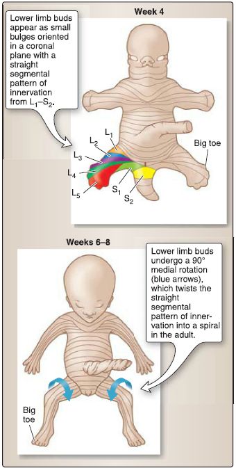
Figure 2: Rotation of the lower limb.
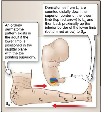
Figure 3: Dermatome pattern of the lower limb.
 الاكثر قراءة في علم التشريح
 اخر الاخبار
اخبار العتبة العباسية المقدسة

الآخبار الصحية


 قسم الشؤون الفكرية يصدر كتاباً يوثق تاريخ السدانة في العتبة العباسية المقدسة
 "المهمة".. إصدار قصصي يوثّق القصص الفائزة في مسابقة فتوى الدفاع المقدسة للقصة القصيرة
 (نوافذ).. إصدار أدبي يوثق القصص الفائزة في مسابقة الإمام العسكري (عليه السلام)


