


النبات

مواضيع عامة في علم النبات
الجذور - السيقان - الأوراق
النباتات الوعائية واللاوعائية
البذور (مغطاة البذور - عاريات البذور)
الطحالب
النباتات الطبية
الحيوان
مواضيع عامة في علم الحيوان
علم التشريح
التنوع الإحيائي
البايلوجيا الخلوية
الأحياء المجهرية
البكتيريا
الفطريات
الطفيليات
الفايروسات
علم الأمراض
الاورام
الامراض الوراثية
الامراض المناعية
الامراض المدارية
اضطرابات الدورة الدموية
مواضيع عامة في علم الامراض
الحشرات
التقانة الإحيائية
مواضيع عامة في التقانة الإحيائية
التقنية الحيوية المكروبية
التقنية الحيوية والميكروبات
الفعاليات الحيوية
وراثة الاحياء المجهرية
تصنيف الاحياء المجهرية
الاحياء المجهرية في الطبيعة
أيض الاجهاد
التقنية الحيوية والبيئة
التقنية الحيوية والطب
التقنية الحيوية والزراعة
التقنية الحيوية والصناعة
التقنية الحيوية والطاقة
البحار والطحالب الصغيرة
عزل البروتين
هندسة الجينات
التقنية الحياتية النانوية
مفاهيم التقنية الحيوية النانوية
التراكيب النانوية والمجاهر المستخدمة في رؤيتها
تصنيع وتخليق المواد النانوية
تطبيقات التقنية النانوية والحيوية النانوية
الرقائق والمتحسسات الحيوية
المصفوفات المجهرية وحاسوب الدنا
اللقاحات
البيئة والتلوث
علم الأجنة
اعضاء التكاثر وتشكل الاعراس
الاخصاب
التشطر
العصيبة وتشكل الجسيدات
تشكل اللواحق الجنينية
تكون المعيدة وظهور الطبقات الجنينية
مقدمة لعلم الاجنة
الأحياء الجزيئي
مواضيع عامة في الاحياء الجزيئي
علم وظائف الأعضاء
الغدد
مواضيع عامة في الغدد
الغدد الصم و هرموناتها
الجسم تحت السريري
الغدة النخامية
الغدة الكظرية
الغدة التناسلية
الغدة الدرقية والجار الدرقية
الغدة البنكرياسية
الغدة الصنوبرية
مواضيع عامة في علم وظائف الاعضاء
الخلية الحيوانية
الجهاز العصبي
أعضاء الحس
الجهاز العضلي
السوائل الجسمية
الجهاز الدوري والليمف
الجهاز التنفسي
الجهاز الهضمي
الجهاز البولي
المضادات الميكروبية
مواضيع عامة في المضادات الميكروبية
مضادات البكتيريا
مضادات الفطريات
مضادات الطفيليات
مضادات الفايروسات
علم الخلية
الوراثة
الأحياء العامة
المناعة
التحليلات المرضية
الكيمياء الحيوية
مواضيع متنوعة أخرى
الانزيمات

Pelvic Osteology
 المؤلف:
Kelly M. Harrell and Ronald Dudek
المؤلف:
Kelly M. Harrell and Ronald Dudek
 المصدر:
Lippincott Illustrated Reviews: Anatomy
المصدر:
Lippincott Illustrated Reviews: Anatomy
 الجزء والصفحة:
 16-7-2021
 2594
2594


+
-
20
Pelvic Osteology
The pelvis is defined as the area and contents contained within the framework of the bony pelvis. Pelvic structures are located between parietal peritoneum of the abdomen and the skin of the perineum. Organs of the genitourinary and distal gastrointestinal (GI) systems make up pelvic viscera.
Unlike the shoulder girdle/complex, the bones of the pelvic girdle form a continuous bony ring that provides for visceral protection and muscle attachment, allows for limited movements, and distributes weight from the vertebral column to the lower limbs.
A. Bony pelvis
The bony pelvis is made up of right and left os coxae anterolaterally and the unpaired sacrum and coccyx posteriorly (Fig. 1 ).
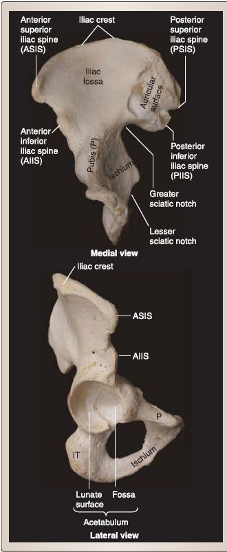
Figure 1 : Osteology of the hip bone.
1. Os coxae (hip bones): Each os coxae is formed by fusion of three bones-ischium, ilium, and pubis. Each bone contributes to the formation of the acetabulum and has important bony landmarks that serve as tendon and ligament attachment sites (Fig. 2).
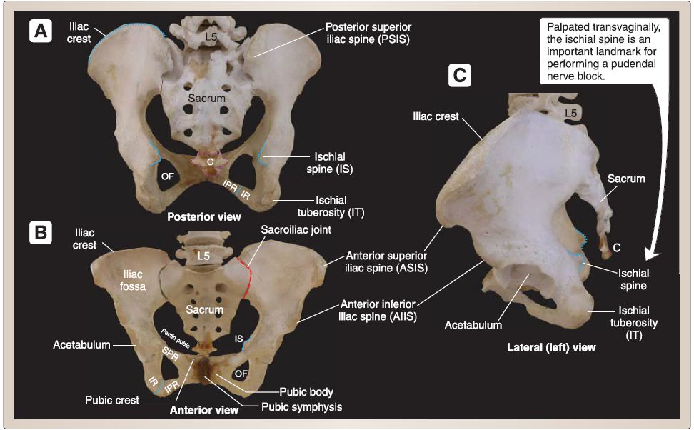 Figure 2: Articulated pelvis. C = coccyx, IPR = inferior pubic ramus, IR = ischial ramus, OF = obturator foramen, SPR = superior pubic ramus.
a. Ilium: The superior lateral portion of the hip is characterized by an anterior concave iliac fossa and a posterior gluteal surface. The superior border of the ilium forms the iliac crest, which terminates anteriorly and posteriorly in projections called anterior superior and posterior superior iliac spines, respectively. Paired anterior and posterior inferior iliac spines lie inferior to their superior counterparts. The medial auricular surface of the ilium articulates with the sacrum posteriorly. The most inferior posterior portion forms the greater sciatic notch.
b. lschium: The posteroinferior portion of the hip is characterized by a large, rough projection of bone, the ischial tuberosity, which serves as an attachment site for hamstring musculature and the sacrotuberous ligament. The ischial spine projects from the superior ischium and is the attachment site for the sacrospinous ligament, superior gemellus, and pelvic floor musculature. lschial rami (superior and inferior) contribute anteriorly to the formation of the obturator foramen.
c. Pubis: The anterior portion of the hip is characterized by a central body and superior and inferior pubic rami, which collectively contribute to the formation of the obturator foramen. The right and left pubic bones articulate anteriorly at the pubic symphysis. Additional important bony landmarks include the pubic tubercle and pectin pubis.
B. Articulations
Joints of the pelvis provide stability, protection, and controlled mobility. As shown in Figure 3, strong ligaments support these joints and, if stretched or injured, can produce pain through malalignment. Joints of the pelvis include the following.
1. Lumbosacral: Articulation of L5 and S1 includes synovial joints between articular processes and a symphysis joint between the L5/S1 intervertebral disc. lliolumbar ligaments (anterior and posterior) support this joint by limiting axial rotation of L5 on S1.
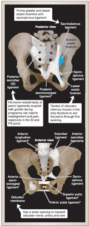
Figure 3:Pelvic ligaments. (Most ligaments are bilateral but illustrated unilaterally here
for simplicity.) PS = pubic symphysis; * = unpaired ligaments.
2. Sacroiliac (SI): Articulations between the right and left auricular surfaces of os coxae and sacrum (right and left SI joints) transmit body weight to the os coxae and the lower limbs and are supported by SI (anterior and posterior) and interosseous ligaments. Secondary support ligaments include the sacrotuberous and sacrospinous ligaments.
a. Greater and lesser sciatic notches: Sacrotuberous and sacrospinous ligaments anchor the sacrum to the ischial tuberosity and ischial spine, respectively. The arrangement of these ligaments turns greater and lesser sciatic notches into greater and lesser sciatic foramina, which serve as passageways for structures to exit and enter the pelvis.
3. Pubic symphysis: This articulation between the right and left pubic bodies is connected through an interpubic fibrocartilage disc and supported by superior and inferior pubic ligaments.
4. Sacrococcygeal: This symphysis articulation between the distal sacrum and coccyx contains a small disc and is supported by sacrococcygeal ligaments.
C. Regions of articulated pelvis
The articulated pelvis can be divided at the level of the pelvic brim into a superior greater (false) pelvis and an inferior lesser (true) pelvis (Fig. 4). The greater pelvis represents the area superior to the pelvic brim that houses abdominal viscera. The lesser pelvis represents the area inferior to the pelvic brim that houses pelvic viscera. The pelvic brim also serves at the boundary for the pelvic inlet (superior pelvic aperture). The pelvic outlet (inferior pelvic aperture) is bound by the coccyx, sacrotuberous ligament, ischial tuberosities, ischiopubic rami, and pubic symphysis.
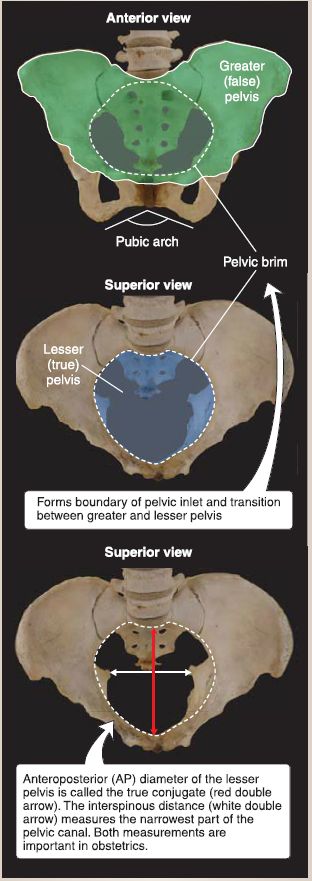
Figure .4:Pelvic regions and boundaries.
 الاكثر قراءة في علم التشريح
 اخر الاخبار
اخبار العتبة العباسية المقدسة

الآخبار الصحية


 قسم الشؤون الفكرية يصدر كتاباً يوثق تاريخ السدانة في العتبة العباسية المقدسة
 "المهمة".. إصدار قصصي يوثّق القصص الفائزة في مسابقة فتوى الدفاع المقدسة للقصة القصيرة
 (نوافذ).. إصدار أدبي يوثق القصص الفائزة في مسابقة الإمام العسكري (عليه السلام)


