Embryology
The CNS (brain and spinal cord) begins to form in week 3 of development during which time the process of neurulation occurs (Fig. 1). Neurulation refers to the formation and closure of the neural tube in which bone morphogenetic protein 4 (BMP-4), noggin (an inductor protein), chordin (an inductor protein), fibroblast growth factor 8, and neural cell adhesion molecule play a role.

Figure 1: Neurulation. The formation and closure of the neural tube. A, Day 20. B, Day 23.
A. Somitomeres
The paraxial mesoderm is a thick plate of mesoderm that organizes into segments called somitomeres, which form in a craniocaudal sequence. The somitomeres one to seven do not form somites but contribute mesoderm to the head and neck region (i.e., the pharyngeal arches). The remaining somitomeres condense in a craniocaudal sequence to form 42-44 pairs of somites of the trunk region. The somites closest to the caudal end eventually disappear, to give a final count of -35 pairs of somites.
1. Somites: The paraxial mesoderm on either side of the mid line (neural tube) gives rise to the somites, which, in turn, soon differentiate into an sclerotome and a dermamyotome (Fig. 2).
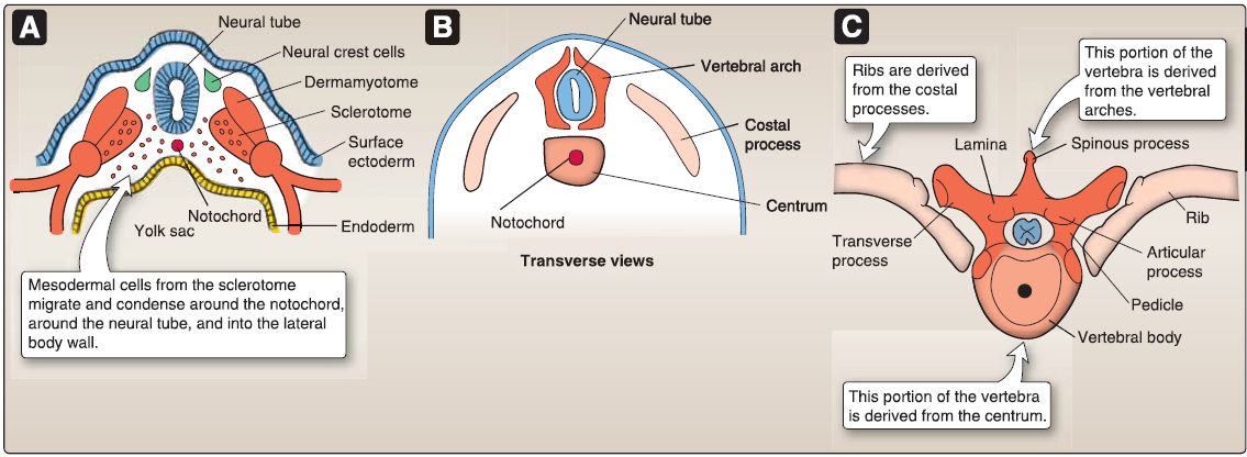 Figure 2: Embryologic formation of vertebrae. A, At about day 28, mesodermal cells from the sclerotome migrate and condense in three areas: around the notochord, around the neural tube, and into the lateral body wall. B, The formation of the centrum, vertebral arches, and the costal processes. C, The adult derivatives of the centrum, vertebral arches, and the costal processes.
a. Sclerotome: During week 4 of development, the mesodermal cells from the sclerotome migrate and condense in three areas: around the notochord, around the neural tube, and into the
lateral body wall.
[1] Sclerodermal cells: The sclerodermal cells around the notochord form the centrum, which develops into the adult vertebral body. The sclerodermal cells around the neural tube form the vertebral arches, which develop into the adult pedicles, laminae, spinous process, articular processes, and the transverse processes. The sclerodermal cells in the lateral body wall form the costal processes, which develop into the adult ribs.
[2] Mesodermal and notochordal cells: The space between the vertebral bodies is filled with mesodermal cells that contribute to the formation of the intervertebral disc. The notochord regresses in the area of the vertebral body, but persists in the intervertebral disc. As mentioned, each intervertebral disc consists of the nucleus pulposus and the annulus fibrosis. The nucleus pulposus is a remnant of the embryonic notochord. By age 20 years, all notochordal cells have degenerated such that all notochordal vestiges in the adult are limited to just a noncellular matrix. The annulus fibrosis is an outer rim of fibrocartilage derived from mesoderm found between the vertebral bodies.
[3] Sclerotome resegmentation: As development continues, each sclerotome splits into a cranial portion with a low cell density and a caudal portion with a high cell density (Fig. 3). The boundary between the cranial portion and the caudal portion is marked by an intersegmental fissure or von Ebner fissure. These fissures provide pathways for the developing spinal nerves to reach their respective myotomes. The caudal portion of each sclerotome fuses with the cranial portion of the sclerotome beneath it, resulting in the intersegmental position of the vertebrae. In the cervical region, the caudal portion of the fourth occipital sclerotome (04) fuses with the cranial portion of the first cervical (C1) sclerotome to form the base of the occipital bone, allowing C1 spinal nerve to exit between the base of the occipital bone and C1 vertebrae.

Figure 3: Resegmentation of the sclerotomes.
b. Dermamyotome: The myotome portion of the dermamyotome forms muscle. The trunk musculature is derived from myotomes in the trunk region. Each myotome partitions into a posterior epimere and an anterior hypomere (Fig. 4).
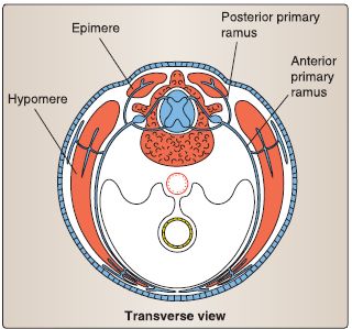
Figure 4:Development of the trunk musculature
[1] Epimere: This develops into the extensor muscles of the neck and vertebral column (e.g., erector spinae). The epimere is innervated by posterior rami of spinal nerves. [2] Hypomere: This develops into the scalene, prevertebral, geniohyoid, infrahyoid, intercostal, and abdominal muscles; lateral and anterior flexors of the vertebral column; the quadratus lumborum; and the pelvic diaphragm. The hypomere is innervated by anterior rami of spinal nerves.
2. Neural plate: Neurulation begins when the primitive node/notochord induces the overlying ectoderm to form the neural plate, which is a thick plate of pseudostratified, columnar neuroepithelial cells called neuroectoderm. The neural plate is shaped with a broad cranial portion that gives rise to the brain and a narrow caudal portion that gives rise to the spinal cord.
3. Neural tube: The neural plate begins to fold during week 4 of development to form a closed neural tube. The closure of the neural tube begins at the future occipital/cervical region and then proceeds in both anterior and posterior directions. This closure continues until the most anterior end (i.e., the anterior neuropore) and the most posterior end (i.e., the posterior neuropore) close. If the anterior neuropore fails to close, upper neural tube defects occur (e.g., anencephaly). If the posterior neuropore fails to close, lower neural tube defects occur (e.g., spina bifida with myeloschisis).As the neural tube folds, some cells differentiate into neural crest cells. The lumen of the neural tube gives rise to the ventricular system of the brain and central canal of the spinal cord.
B. Special development zones
The early neural tube consists of neuroectoderm arranged in three zones called the ventricular zone, intermediate zone, and marginal zone (Fig.5).
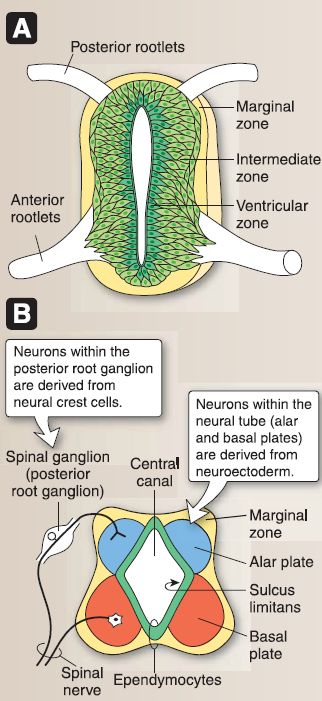
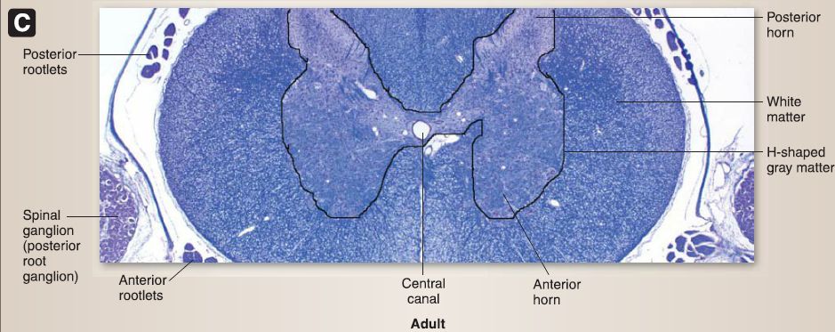 Figure 5: A and B, Further development of the neural tube. C, Posterior and anterior horns of the adult spinal cord.
1. Ventricular zone: Two waves of proliferation/differentiation occur here. The first wave gives rise to neuroblasts, which migrate into the intermediate zone. The second wave gives rise to glioblasts, which migrate into the intermediate and marginal zones. The remaining neuroectoderm gives rise to the ependymocytes that line the central canal of the spinal cord.
2. Intermediate zone: After migration, the neuroblasts differentiate into neurons, and the glioblasts differentiate into astrocytes and oligodendrocytes. The intermediate zone forms the H-shaped gray matter of the spinal cord, which is divided into the alar plate and the basal plate. The alar plate becomes the posterior horn of the spinal cord. The basal plate becomes the anterior horn of the spinal cord. Both the alar plate and the basal plate are separated by a longitudinal groove called the sulcus limitans.
3. Marginal zone: The marginal zone contains axons that arise from the neurons within the intermediate zone. In addition, the marginal zone contains glioblasts that differentiate into astrocytes and oligodendrocytes. The marginal zone forms the white matter of the spinal cord.
C. Positional changes
The position of the spinal cord in relationship to the vertebral column changes due to the disparate growth between the spinal cord and vertebral column (Fig. 2.27). At week 8 of development, the spinal cord extends the length of the vertebral canal. At birth, the conus medullaris (the tapered lower end of the spinal cord) extends to the level of the third lumbar vertebra (L3). In adults, the conus medullaris terminates at the L 1-L2 interspace. The disparate growth also results in the formation of the cauda equina that consists of the posterior and anterior roots from L3 to coccygeal vertebral levels and that descends below the level of the conus medullaris. Additionally, this disparate growth results in the formation of the non-neural filum terminale (pia), which anchors the spinal cord inferiorly to the coccyx. D. Neural crest cell formation Neural crest cells differentiate from cells located along the lateral border of the neural plate, which is mediated by BMP-4 and BMP-7.
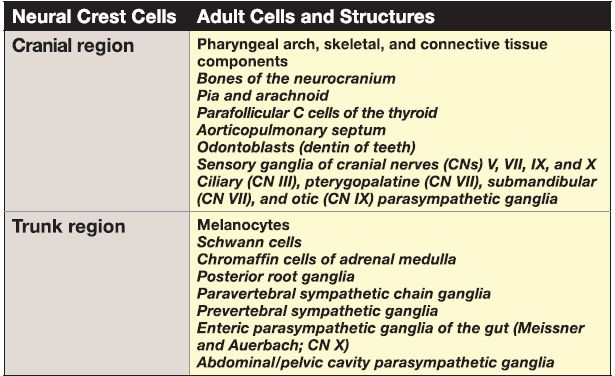
They can be generally divided into cranial neural crest cells and trunk neural crest cells based on their prolific migration throughout the embryo to both the cranial and trunk regions. Neural crest cells ultimately differentiate into a wide array of adult cells and structures, as indicated in Table 1.
Table 1: Adult Cells and Structures Formed From Neural Crest Cells