


النبات

مواضيع عامة في علم النبات
الجذور - السيقان - الأوراق
النباتات الوعائية واللاوعائية
البذور (مغطاة البذور - عاريات البذور)
الطحالب
النباتات الطبية
الحيوان
مواضيع عامة في علم الحيوان
علم التشريح
التنوع الإحيائي
البايلوجيا الخلوية
الأحياء المجهرية
البكتيريا
الفطريات
الطفيليات
الفايروسات
علم الأمراض
الاورام
الامراض الوراثية
الامراض المناعية
الامراض المدارية
اضطرابات الدورة الدموية
مواضيع عامة في علم الامراض
الحشرات
التقانة الإحيائية
مواضيع عامة في التقانة الإحيائية
التقنية الحيوية المكروبية
التقنية الحيوية والميكروبات
الفعاليات الحيوية
وراثة الاحياء المجهرية
تصنيف الاحياء المجهرية
الاحياء المجهرية في الطبيعة
أيض الاجهاد
التقنية الحيوية والبيئة
التقنية الحيوية والطب
التقنية الحيوية والزراعة
التقنية الحيوية والصناعة
التقنية الحيوية والطاقة
البحار والطحالب الصغيرة
عزل البروتين
هندسة الجينات
التقنية الحياتية النانوية
مفاهيم التقنية الحيوية النانوية
التراكيب النانوية والمجاهر المستخدمة في رؤيتها
تصنيع وتخليق المواد النانوية
تطبيقات التقنية النانوية والحيوية النانوية
الرقائق والمتحسسات الحيوية
المصفوفات المجهرية وحاسوب الدنا
اللقاحات
البيئة والتلوث
علم الأجنة
اعضاء التكاثر وتشكل الاعراس
الاخصاب
التشطر
العصيبة وتشكل الجسيدات
تشكل اللواحق الجنينية
تكون المعيدة وظهور الطبقات الجنينية
مقدمة لعلم الاجنة
الأحياء الجزيئي
مواضيع عامة في الاحياء الجزيئي
علم وظائف الأعضاء
الغدد
مواضيع عامة في الغدد
الغدد الصم و هرموناتها
الجسم تحت السريري
الغدة النخامية
الغدة الكظرية
الغدة التناسلية
الغدة الدرقية والجار الدرقية
الغدة البنكرياسية
الغدة الصنوبرية
مواضيع عامة في علم وظائف الاعضاء
الخلية الحيوانية
الجهاز العصبي
أعضاء الحس
الجهاز العضلي
السوائل الجسمية
الجهاز الدوري والليمف
الجهاز التنفسي
الجهاز الهضمي
الجهاز البولي
المضادات الميكروبية
مواضيع عامة في المضادات الميكروبية
مضادات البكتيريا
مضادات الفطريات
مضادات الطفيليات
مضادات الفايروسات
علم الخلية
الوراثة
الأحياء العامة
المناعة
التحليلات المرضية
الكيمياء الحيوية
مواضيع متنوعة أخرى
الانزيمات

Spinal Cord
 المؤلف:
Kelly M. Harrell and Ronald Dudek
المؤلف:
Kelly M. Harrell and Ronald Dudek
 المصدر:
Lippincott Illustrated Reviews: Anatomy
المصدر:
Lippincott Illustrated Reviews: Anatomy
 الجزء والصفحة:
 10-7-2021
 5544
5544


+
-
20
Spinal Cord
Housed and protected within the vertebral column, the spinal cord spans from the foramen magnum to approximately the L2 vertebral level in the adult (Fig. 1). As part of the central nervous system (CNS), the spinal cord serves as a reflex center and conducts motor and sensory signals between the brain and the body. The vertebral column also contains vasculature, adipose, and meningeal layers, which supply and protect the spinal cord, respectively. The spinal cord is cylindrical in shape with enlargements in the cervical and lumbar regions to accommodate for upper and lower limb innervation, respectively. Distally, the spinal cord tapers and ends at L2 as the conus medullaris. Multiple spinal nerve rootlets emerge from the distal spinal cord and conus medullaris to exit the vertebral column. They are collectively referred to as the cauda equina .
A. Internal organization
As shown in Figure 2, in cross section, the spinal cord has an "H"-shaped gray matter core made up of neuronal cell bodies and peripheral white matter made up of nerve fibers (axons).
1. Gray matter: Gray matter is organized into anterior and posterior horns, which contain motor and sensory cell bodies, respectively. The thoracolumbar region {T1-L/L3) also features a lateral horn that contains preganglionic sympathetic cell bodies organized into an intermediolateral cell column.
2. White matter: Peripheral white matter is organized into specific ascending (sensory) and descending (motor) tracts.
B. Spinal nerves
Spinal nerves carry autonomic and somatic motor (general visceral efferent [GVE] and general somatic efferent [GSE]) and sensory (general visceral afferent [GVA] and general somatic afferent [GSA]) nerve fibers. Thirty-one paired spinal nerves exit the vertebral column through the intervertebral foramina or sacral foramina at each vertebral level (8 cervical, 12 thoracic, 5 lumbar, 5 sacral, and 1 coccygeal).
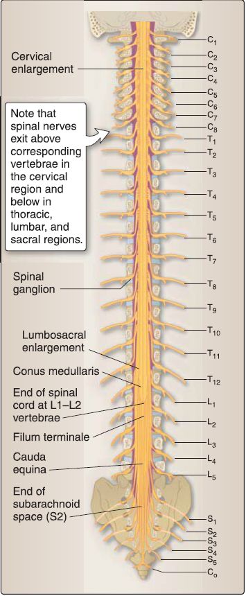
Figure 1:Spinal cord. Posterior schematic image of the spinal cord within the vertebral column (laminae removed) and exiting spinal nerves.
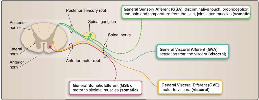
Figure 2: Cross section of the spinal cord with spinal nerve components.
1. Anterior and posterior rootlets: These arise from the spinal cord laterally, representing motor and sensory fibers, respectively (see Fig. 2). Rootlets converge to form nerve roots, which contain autonomic and somatic motor (anterior root) or sensory (posterior root) components.
2. Pseudounipolar sensory neurons: Cell bodies for these are housed in the spinal ganglion (posterior root ganglion) that is contained in the posterior root (Fig. 3).
3. Multipolar motor neurons: Cell bodies for these are found in the anterior horn of the spinal cord, and their axons form the anterior root.
4. Rami: Once the spinal nerve has exited the vertebral column, it splits into an anterior and a posterior ramus. Anterior rami provide motor and sensory innervation to the skin, muscles, vasculature, and joints of the trunk and extremities, while the posterior rami innervate intrinsic back muscles, associated vasculature, and overlying skin.

Figure 3 :Cadaveric image showing caudal spinal cord structures {laminae removed).
C. Meninges
Spinal meninges include the dura mater, arachnoid mater, and pia mater, which collectively surround, support, and protect the spinal cord and spinal nerve roots (Fig. 4).
1. Dura mater: The dura mater (Latin for tough mother) is the outermost layer and is a thick, tough covering. An epidural (extradural) space separates the dura from the boundaries of the vertebral foramen that is filled with adipose and a spinal venous plexus. Spinal dura mater is continuous with the cranial dura mater and extends inferiorly to the level of S2, forming a tube-like dural sac (Fig. 4).
2. Arachnoid mater: Deep to the dura mater is the arachnoid mater, a delicate, web-like layer that creates a subarachnoid space filled with cerebrospinal fluid (CSF). CSF bathes and protects the spinal cord and nerve roots within the vertebral canal. The subarachnoid space also contains arterial and venous branches that nourish the spinal cord. Under normal conditions, the arachnoid and dura are closely associated, obliterating the subdural space between these two layers. However, bleeding can occur into this space, forming a subdural hematoma. A widened subarachnoid space exists below the level of the conus medullaris between vertebral levels L2-S2. This space, referred to as the lumbar cistern, lies within the dural sac and contains CSF and the cauda equina.
3. Pia mater: The innermost meningeal layer, the pia mater, is closely adhered to the spinal cord. Two specializations of pia mater function to anchor the spinal cord within the vertebral canal: Denticulate ligaments are extensions of pia that project laterally at each vertebral foramen level and anchor into the inner surface of the dura mater, separating anterior and posterior rootlets (Fig. 5). The filum terminale is an extension of pia from the tip of the conus medullaris that extends inferiorly to attach on the coccyx, acting as a caudal anchor for the spinal cord.
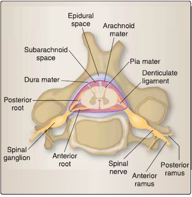
Figure4 : Cross-sectional view of spinal meninges and spaces.
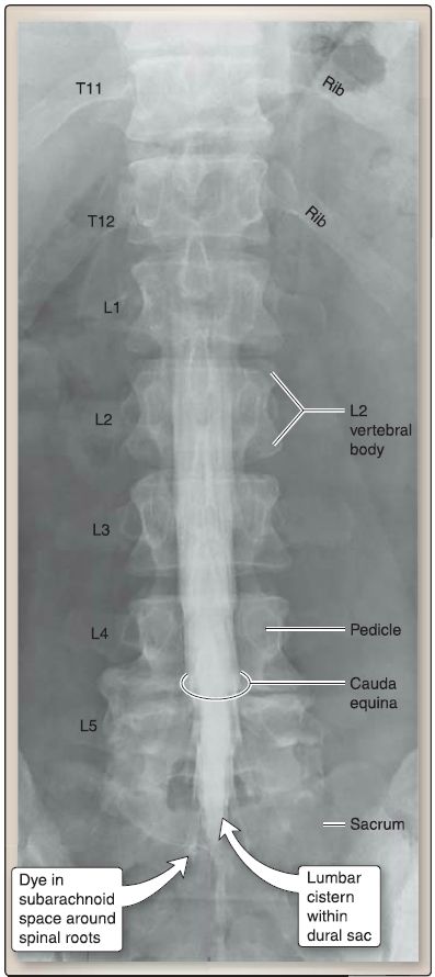
Figure 5 : Lumbosacral myelography.
 الاكثر قراءة في علم التشريح
 اخر الاخبار
اخبار العتبة العباسية المقدسة

الآخبار الصحية


 قسم الشؤون الفكرية يصدر كتاباً يوثق تاريخ السدانة في العتبة العباسية المقدسة
 "المهمة".. إصدار قصصي يوثّق القصص الفائزة في مسابقة فتوى الدفاع المقدسة للقصة القصيرة
 (نوافذ).. إصدار أدبي يوثق القصص الفائزة في مسابقة الإمام العسكري (عليه السلام)


