آخر المواضيع المضافة

 النبات
الحيوان
الأحياء المجهرية
علم الأمراض
التقانة الإحيائية
التقنية الحيوية المكروبية
التقنية الحياتية النانوية
علم الأجنة
الأحياء الجزيئي
علم وظائف الأعضاء
الغدد
المضادات الحيوية
النبات
الحيوان
الأحياء المجهرية
علم الأمراض
التقانة الإحيائية
التقنية الحيوية المكروبية
التقنية الحياتية النانوية
علم الأجنة
الأحياء الجزيئي
علم وظائف الأعضاء
الغدد
المضادات الحيوية| Spleen |


|
|
Read More
Date: 23-1-2017
Date: 10-1-2017
Date: 27-7-2016
|
Spleen
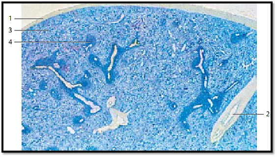
The spleen is the largest lymphatic organ in the human body. It is enveloped by a collagen fiber capsule 1 , which has a covering of peritoneal epithelium (top of the figure). Irregularly arrange d connective tissue cords traverse the organ, starting at the outer capsule. They are named splenic trabeculae 2 (almost unstained in this preparation) and contain the trabecular blood vessels. Richly vascularized reticular connective tissue occupies the space between splenic capsule and trabeculae. The abundance of blood explains the name re d spleen pulp 3 for the flexible reticular web. The pulp contains splenic follicles 4 (stained darker blue), which in their entirety represent the white pulp . This white pulp forms a reticular tissue with many lymphocytes, which surrounds the vessels like a sheath. Many of these vessels are cut longitudinally in this preparation.
1 Capsule
2 Spleen trabeculae with trabecular vein
3 Red splenic pulp
4 Splenic nodule with central artery
Stain: alum hematoxylin; magnification: × 4
Spleen
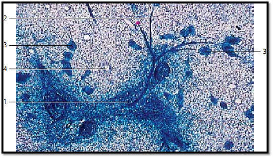
This section shows part of a feline spleen with a splenic follicle (spleen follicle; periarterial lymphocyte sheath ), which contains the follicular or central artery 1 . The central artery splits into 25–50 arterioles, sometimes still inside the follicle, but mostly at the exit point. The arterioles spread out like a brush or tree (brush arterioles , penicillus) 2 . The penicillar arterioles are covered by a spindle-shape d sheath 3 (Schweigger-Seidel sheath, ellipsoid) shortly after they have split off the central artery (sheath arterioles). The sleeve-like sheaths are hard to find in a human spleen. However, in a feline spleen they appear as prominent small follicles. They consist of dense reticular connective tissue and macrophages. The lighter stained and loosely structured areas are part of the re d splenic pulp 4 . The feline spleen was perfused with physiological saline solution before specimen preparation.
1 Splenic follicle
2 Brush arteriole
3 Sleeve-like sheath
4 Red splenic pulp
Stain: methyl blue; magnification: × 10
Spleen
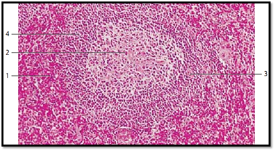
The parenchyme of the spleen consists of the white pulp (= all lymphoreticular arterial sheaths, T-cell region ) and the red pulp 1 . This figure shows white pulp as part of a round lymph follicle (folliculus lymphaticus splenicus ). It features a prominent germinal center 2 . The branches of the trabecular artery run through the follicle and are now called follicular artery 3 or central artery. There are usually two to three follicular arteries. They traverse the arterial sheaths eccentrically, especially when secondary follicles with germinal centers (B-cell regions) are present, as is the case here. A barrier of small lymphocytes surrounds the germinal center 2 , which is called corona or mantle zone 4 . The area around the splenic follicle represents re d pulp 1.
1 Splenic red pulp
2 Germinal center of a spleen follicle
3 Follicular (central) artery
4 Corona or mantle zone
Stain: alum hematoxylin-eosin; magnification: × 100
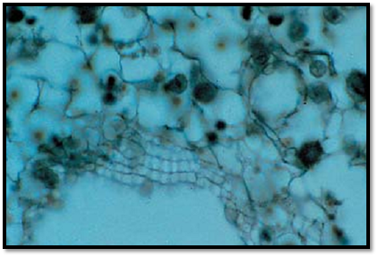
Spleen
From follicular arterioles (penicillar arterioles) derive terminal capillaries, which continue in the surrounding splenic sinuses, except the marginal sinus. There, they supply anastomosing venous sinuses. Terminal capillaries can only be studied in the perfused spleen. The wall of a splenic sinus is built in the form of a grid. It consists of elongated sinus endothelium and argyrophilic fibers in a circular arrangement. The figure provides a view of the surface of the reticular connective tissue. The typical network can be seen in the top half of the figure.
Stain: alum hematoxylin; magnification: × 800
Spleen
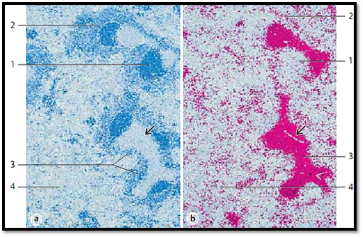
This figure shows the distribution of B- and T-lymphocytes in the spleen. As shown by immunohistochemistry, B-lymphocytes (stained blue) are located mostly in follicles 1 and the marginal zone 1 . The lymphatic arterial sheath 3 , that is pre dominantly the area around the central artery, contains only few B-lymphocytes . The section following the one described in (a) shows the localization of T-cell receptors (red). They are found in large numbers in the lymphatic arterial sheaths 3 . Apart from many macrophages, there are also B- and T-lymphocytes in the splenic red pulp 4 .
1 Follicles
2 Marginal zone
3 Lymphatic arterial sheath
4 Red pulp. The darts show the central arteries.
Magnification: × 60
References
Kuehnel, W.(2003). Color Atlas of Cytology, Histology, and Microscopic Anatomy. 4th edition . Institute of Anatomy Universitätzu Luebeck Luebeck, Germany . Thieme Stuttgart · New York .


|
|
|
|
التوتر والسرطان.. علماء يحذرون من "صلة خطيرة"
|
|
|
|
|
|
|
مرآة السيارة: مدى دقة عكسها للصورة الصحيحة
|
|
|
|
|
|
|
نحو شراكة وطنية متكاملة.. الأمين العام للعتبة الحسينية يبحث مع وكيل وزارة الخارجية آفاق التعاون المؤسسي
|
|
|