آخر المواضيع المضافة

 النبات
الحيوان
الأحياء المجهرية
علم الأمراض
التقانة الإحيائية
التقنية الحيوية المكروبية
التقنية الحياتية النانوية
علم الأجنة
الأحياء الجزيئي
علم وظائف الأعضاء
الغدد
المضادات الحيوية
النبات
الحيوان
الأحياء المجهرية
علم الأمراض
التقانة الإحيائية
التقنية الحيوية المكروبية
التقنية الحياتية النانوية
علم الأجنة
الأحياء الجزيئي
علم وظائف الأعضاء
الغدد
المضادات الحيوية| Lymph Nodes |


|
|
Read More
Date: 27-7-2016
Date: 25-7-2016
Date: 15-1-2017
|
Lymph Nodes
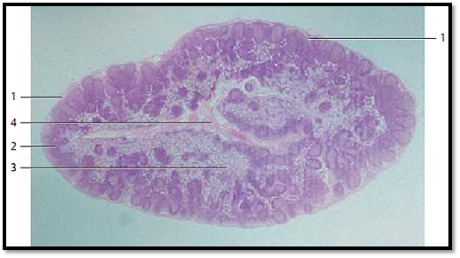
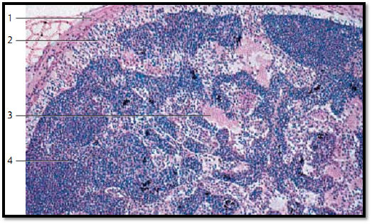
Lymph nodes, nodi lymphatici , are oval or bean-shaped organs. They function as biological filters in the lymphatic circulation. Their sizes may range b e-tween several millimeters to more than 2 centimeters. They are encapsulate d by connective tissue collagen fibers 1 . Connective tissue divisions from the capsule, the trabeculae, extend into the interior node. Sporadic myocytes are found inside the capsule. Trabeculae form the basic skeleton of the organ. The finer network of reticulum cells and the argyrophilic fibers (reticular fibers) is interspersed with this skeleton. The web contains considerable numbers of lymphocytes and macrophages (lymphoreticular organ ). Lymph nodes consist of the cortex 2 and inner medulla 3 . Blood vessels enter at the hilus region, and efferent lymph vessels (vasa efferentia) from the lymph node exit at the hilus. Several afferent lymph vessels (vasa afferentia) pierce the capsule and bring lymph into the lymph node. The figure very clearly shows the heavily stained cortex and the lighter medulla. In the cortex, we see a large number of nodular follicles with germinal centers ( lymphatic nodules). Please, note the central medullary sinus 4. Human inguinal lymphatic node.
1 Capsule
2 Corte x with secondary follicles (lymphatic nodules)
3 Medulla
4 Medullary sinus
Stain: alum hematoxylin-eosin; magnification: × 20
Lymph Nodes
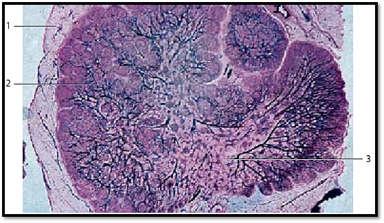
A small lymph node from a rabbit was injected with Indian ink . It is possible to clearly recognize the strong connective tissue capsule 1 , the heavily stained cortex 2 with lymphatic nodules and the lighter medulla 3 as well as the finer branches of the arteries, which extend from the hilus region to the trabeculae, the medulla and, finally, to the cortex.
1 Capsule
2 Corte x
3 Medulla
Tissue injection with Indian ink; stain: hemalum-eosin; magnification: × 5
Lymph Nodes
Section of a lymph node. The surface is encased in a strong connective tissue capsule 1 . Lymph vessels close to the lymph nodes (vasa afferentia) pierce this capsule and enter the outer cortical sinus (sinus marginalis) 2 , which is clearly accentuated as a lighter crevice between capsule and cortex with only a few lymphocytes. The marginal sinus is connected to the radial structure of the trabecular sinus, which ultimately leads to the central medullary sinus 3 with its wider lumen. Lymphocyte-rich areas, named primary noduli, populate the cortex 4 . The small piles of darker granules are carbon inclusions.
1 Capsule
2 Marginal sinus
3 Medullary sinus
4 Cortex
Stain: hemalumeosin; magnification × 20
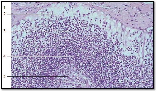
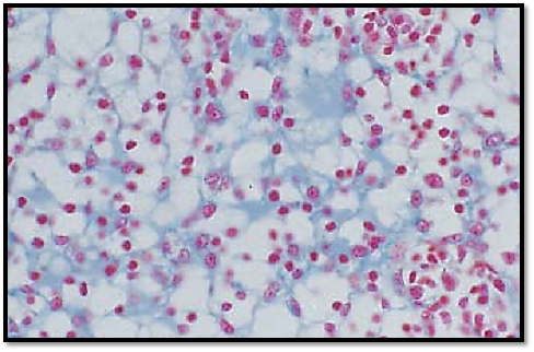
Lymph Nodes
Outer region of a lymph node from the human axilla, with a relatively tough connective tissue capsule 1 , marginal sinus 2 and cortex 3. Reticulum cells (sinus reticulum ) and reticulum f ibers are clearly visible in the marginal sinus . They traverse the marginal sinus and form a loose meshwork . Note the sub capsular cells, name d mantle cells, which are similar to endothelial cells. Intermediary sinuses, which push through the compact cortical tissue, connect the marginal sinuses with the wide medullary sinuses. Lymphocytes are densely packed underneath the marginal sinus. They form a secondary follicle with a lymph corona 4 and a germinal center 5 .
1 Capsule
2 Marginal sinus
3 Cortex
4 Lymph corona of the secondary follicle
5 Germinal center
Stain: alum hematoxylin-eosin; magnification: × 200
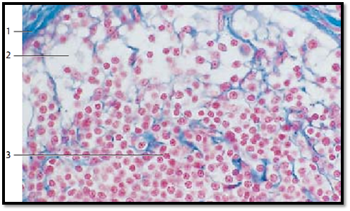
Lymph Nodes
Outer zone of a human inguinal lymph node. The marginal sinus ( sinus marginalis) wraps around the lymph node immediately underneath the capsule 1 like a shell. Numerous afferent lymph vessels ( vasa afferentia) discharge into this marginal sinus. Reticulum cells with many cytoplasmic processes span the marginal sinus. Delicate collagen and reticulum fibers (here stained light blue) lend mechanic support to the cells. A follicle with many lymphocytes (a lymphatic nodule ) 3 is shown in the lower half of the figure.
1 Capsule
2 Marginal sinus
3 Lymph follicle
Stain: azan; magnification: × 400
Lymph Nodes
The lymphocytes in the outer layer of the cortex occur condense d in ovoid or round nodes, called follicles or noduli lymphatici . These follicles are the B-lymphocyte regions of the lymph node. The regions with T-lymphocytes are the diffusely delimited paracortical zones. Cortical nodes solely consist of densely packed small and me diumsized lymphocytes. They are called primary follicles. This preparation presents a secondary follicle. Apart from small lymphocytes, it contains in its center large cells with basophilic cytoplasm (centrocytes and centroblasts). These regions inside a follicle are called germinal centers 1 . They arise from primary follicles during immunogenesis. A lymphocyte barrier (corona) 2 surrounds the germinal center of a secondary follicle. It pre dominantly consists of small lymphocytes. Sporadically, plasma cells occur in the lymphocyte corona.
1 Germinal center
2 Lymphocyte corona (barrier)
Stain: azan; magnification: × 200
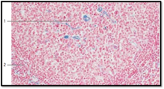
Lymph Nodes
Section of a medullary sinus from a feline lymph node. The medullary sinuses are located between medullary cords. They connect to the marginal sinus via intermediary sinus. Reticulum cells traverse the medullary sinus as well as the other lymph node sinuses. This creates a bow net system. Note the lacy web of the reticulum cells (stained light blue). Their structure is sponge-like. Lymphocytes, of ten macrophages, occasionally monocytes and plasma cells occur in the mesh-work .
Stain: azan; magnification: × 400
References
Kuehnel, W.(2003). Color Atlas of Cytology, Histology, and Microscopic Anatomy. 4th edition . Institute of Anatomy Universitätzu Luebeck Luebeck, Germany . Thieme Stuttgart · New York .


|
|
|
|
لخفض ضغط الدم.. دراسة تحدد "تمارين مهمة"
|
|
|
|
|
|
|
طال انتظارها.. ميزة جديدة من "واتساب" تعزز الخصوصية
|
|
|
|
|
|
|
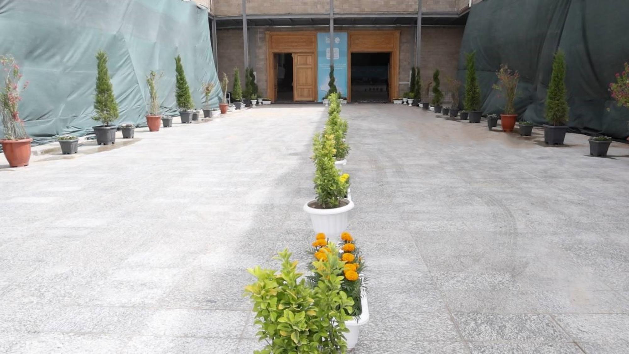
مشاتل الكفيل تزيّن مجمّع أبي الفضل العبّاس (عليه السلام) بالورد استعدادًا لحفل التخرج المركزي
|
|
|