آخر المواضيع المضافة

 النبات
الحيوان
الأحياء المجهرية
علم الأمراض
التقانة الإحيائية
التقنية الحيوية المكروبية
التقنية الحياتية النانوية
علم الأجنة
الأحياء الجزيئي
علم وظائف الأعضاء
الغدد
المضادات الحيوية
النبات
الحيوان
الأحياء المجهرية
علم الأمراض
التقانة الإحيائية
التقنية الحيوية المكروبية
التقنية الحياتية النانوية
علم الأجنة
الأحياء الجزيئي
علم وظائف الأعضاء
الغدد
المضادات الحيوية| Kinocilia-Tuba Uterina |


|
|
Read More
Date: 14-8-2016
Date: 14-8-2016
Date: 31-7-2016
|
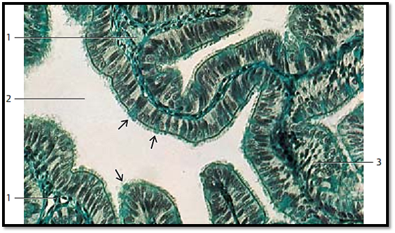
Kinocilia-Tuba Uterina
Kinocilia are motile, membrane-enclosed cell processes. They emerge from small bodies ( basal bodies or kinetosomes) under the cell membrane. Cilia are usually 2–5 μm long and have diameters of about 0.2–0.3 μm. That makes them considerably longer than microvilli and easily discernible using light microscopy. Kinocilia are often abundant (cilia border) and form very dense groups at the cell surface. Such cells are called cilia cells.
This figure shows single-layered columnar epithelial cells from the oviduct mucosa, which consists of cilia cells and secretory cells . As seen in light microscopy, the cilia originate with the row of heavily stained basal bodies ( line of basal bodies ). The secretory cells protrude like domes into the lumen of the duct . They interrupt the rows of basal bodies at the inner plasmalemma .
1 Loosely organized connective tissue of the mucosa folds
2 Oviduct lumen
3 Horizontal or angular section through oviduct epithelium
Stain: Masson-Goldner trichrome stain; magnification: × 200
References
Kuehnel, W.(2003). Color Atlas of Cytology, Histology, and Microscopic Anatomy. 4th edition . Institute of Anatomy Universitätzu Luebeck Luebeck, Germany . Thieme Stuttgart · New York .


|
|
|
|
اكتشاف تأثير صحي مزدوج لتلوث الهواء على البالغين في منتصف العمر
|
|
|
|
|
|
|
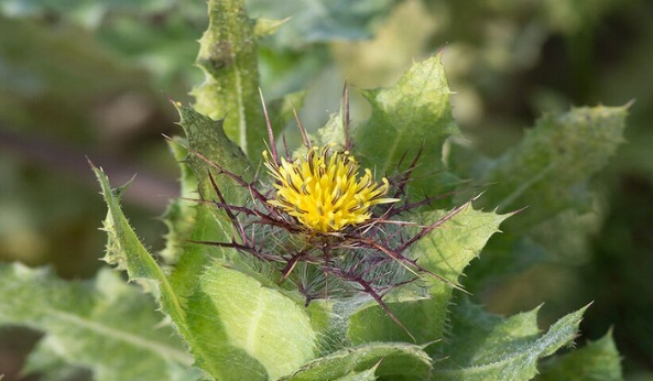
زهور برية شائعة لتر ميم الأعصاب التالفة
|
|
|
|
|
|
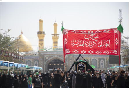
موكب أهالي كربلاء يستذكر شهادة الإمام الصادق (عليه السلام)
|
|
|
|
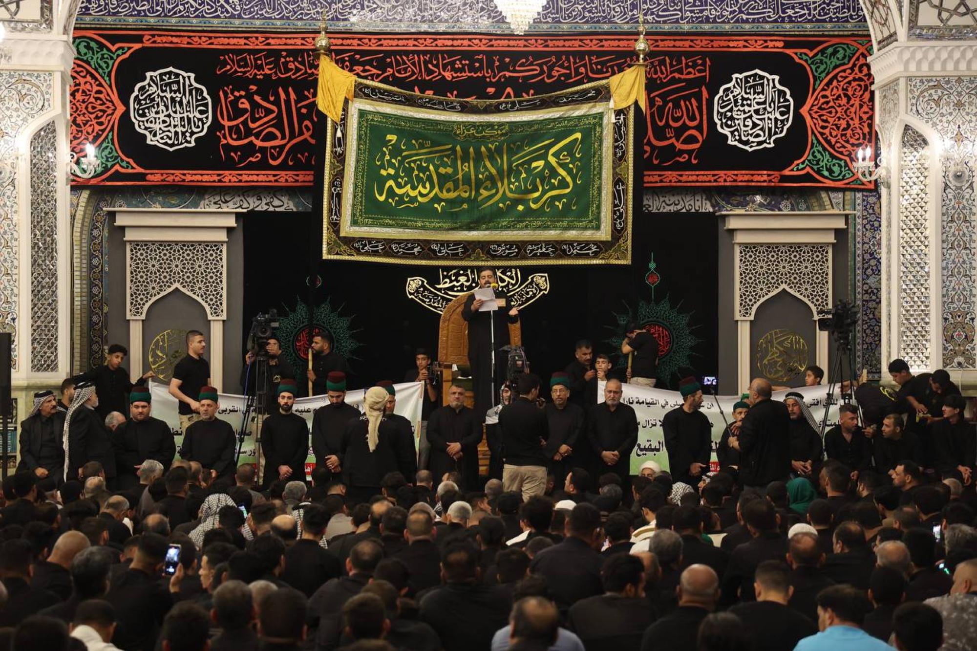
العتبة العباسية تستذكر شهادة الإمام الصادق (عليه السلام) بإقامة مجلس عزاء
|
|
|
|
أهالي كربلاء يحيون ذكرى شهادة الإمام الصادق (عليه السلام) في مدينة الكاظمية
|
|
|
|
شعبة مدارس الكفيل النسوية تعقد اجتماعًا تحضيريًّا لوضع الأسئلة الامتحانية
|