آخر المواضيع المضافة

 النبات
الحيوان
الأحياء المجهرية
علم الأمراض
التقانة الإحيائية
التقنية الحيوية المكروبية
التقنية الحياتية النانوية
علم الأجنة
الأحياء الجزيئي
علم وظائف الأعضاء
الغدد
المضادات الحيوية
النبات
الحيوان
الأحياء المجهرية
علم الأمراض
التقانة الإحيائية
التقنية الحيوية المكروبية
التقنية الحياتية النانوية
علم الأجنة
الأحياء الجزيئي
علم وظائف الأعضاء
الغدد
المضادات الحيوية| Petrifilm™ AOAC official method 2003.1 for Enterobacteriaceae in selected foods |


|
|
Read More
Date: 8-3-2016
Date: 18-3-2016
Date: 14-3-2016
|
Petrifilm™ AOAC official method 2003.1 for Enterobacteriaceae in selected foods
Official method of the AOAC International, as described in the Revision 3 of the 18th Edition of the Official Methods of Analysis of AOAC International (Horwitz and Latimer, 2010). Applied to milk, cheddar cheese, flour, frozen prepared meals, frozen broccoli, and nut pieces.
Petrifilm™ (3M Company) is a modified version of the Colony Forming Units (CFU) plate count method, consisting of two sterile, dry rehydratable films impregnated with culture medium and cold-water-soluble gel-ling agents. Inoculation is done on the surface of the plating surface (bottom film), which, after inoculation is covered with the top film. The inoculum is spread evenly using a plastic spreader and applying gentle manual pressure. After solidification of the gel, the plates are incubated for the development of colonies. The culture medium is Violet Red Bile Glucose (VRBG) Agar supplemented with 2,3,5-triphenyltetrazolium chloride (an indicator which, when reduced, gives the colonies a deep red color) and with an pH indicator (to detect fermentation with acid production, with or without the production of gas). Typical Enterobacteriaceae colonies are red, surrounded by a yellow halo with or without gas bubbles.
1 - Material required for analysis
Preparation of the sample and serial dilutions
• Diluent: 0.1% Peptone Water (PW) or Butterfield’s Phosphate Buffer
• Dilution tubes containing 9 ml 0.1% Peptone Water (PW) or Butterfield’s Phosphate Buffer
Inoculation and incubation
• Petrifilm Enterobacteriaceae plates
• Laboratory incubator set to 35 ± 1°C
2 - Procedure
a) Preparation of the samples and serial dilutions.
Do not use diluents containing citrate, bisulphite or thiosulphate with Petrifilm plates as they can inhibit growth. In those cases in which these diluents, replace by Butterfield’s Phosphate Buffer. After homogenization, withdraw an aliquot of known volume and check the pH. If necessary, adjust the pH to the 6.5–7.5 range with NaOH or HCl 1N, and determine and record the volume of acid or base consumed in the adjustment. Add to the sample a proportional volume of sterile NaOH or HCl solution.
b) Inoculation. Select three appropriate dilutions of the sample and inoculate 1 ml of each dilution on a Petrifilm Enterobacteriaceae plate. To that purpose, follow the manufacturer’s instructions, i.e.: Place the plate on a flat, level surface, lift the top film and place the tip of the pipette perpendicular to the center of the bottom film. Dispense 1 ml on the plating surface and cover the liquid with the top film, taking care to avoid the formation of bubbles. Place the plastic spreader with the recessed side down on the center of the plate. Press gently on the center of the spreader to distribute the sample evenly over the entire Petrifilm plate growth area. Do not drag the spreader over the plate surface, just press gently. Remove the spreader and leave the plate undisturbed for two to five minutes, to permit the gel to form and until complete solidification.
Note b.1) To select dilutions, follow the instructions contained in Chapter 3, since Petrifilm plate count is based on the same principles as the traditional plate count method.
Note b.2) Some foods may interfere with visualization of colonies in first dilution, such as is the case with coffee, chocolate or dried herbs (very dark).
c) Incubation. Incubate the plates at 35 ± 1°C/ 24 ± 2 h, with the clear side up in stacks of no more than 20 plates.
Note c.1) It may be necessary to humidify the incubator to prevent over-drying and dehydrating the plates. Moisture loss, as indicated by weight loss, should not exceed 15% after incubation.
d) Counting the colonies and calculating the results. Select plates with 15 to 100 colonies. Count only typical colonies, which can be of three types: red with gas bubbles and without yellow halo, red with yellow halo but without gas bubbles or red with yellow halo and with gas bubbles. Determine the number of colony-forming units CFU/g or ml by multiplying the number of typical colonies by the inverse of dilution (CFU/g or ml = No colonies/dilution).
Note d.1) Do not count the colonies present in the foam barrier at the edge of the film, since these did not undergo the action of the selective agents. Do not enumerate artificial air bubbles.
Note d.2) The circular growth area of the Petrifilm plates is about 20 cm2 in size. On plates with more than 150 colonies, counts may be estimated by counting the number of colonies in one or more representative 1-cm square areas and calculating the average per 1-cm square area. Next, multiply this number by 20 to obtain the total number of colonies per plate.
Note d.3) After the incubation time the plates may be stored frozen (at −15°C or lower temperatures) for counting at a later time.
References
Silva, N.D .; Taniwaki, M.H. ; Junqueira, V.C.A.; Silveira, N.F.A. , Nasdcimento , M.D.D. and Gomes ,R.A.R .(2013) . Microbiological examination methods of food and water a laboratory Manual. Institute of Food Technology – ITAL, Campinas, SP, Brazil .
Horwitz, W. & Latimer, G.W. (eds) (2010) Official Methods of Analysis of AOAC International. 18th edition, revision 3. Gaithersburg, Maryland, AOAC International.


|
|
|
|
كيف تعزز نمو الشعر الصحي؟
|
|
|
|
|
|
|
10 فحوصات مهمة يجب القيام بها لسيارتك قبل الصيف
|
|
|
|
|
|
جامعة الزهراء (عليها السلام) تكرم قسم الشؤون الفكرية بمناسبة اليوم العالمي للكتاب
|
|
|
|
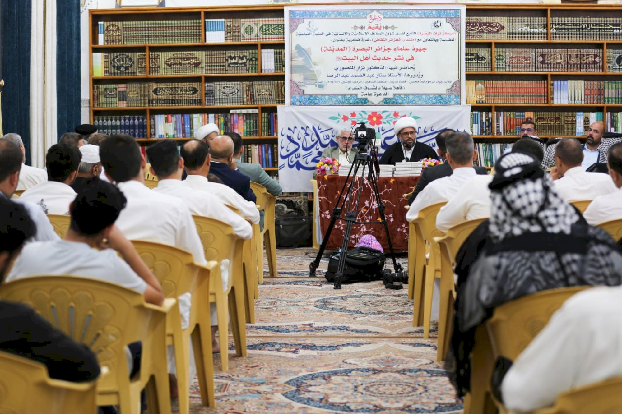
قسم شؤون المعارف يقيم ندوة علمية حول جهود علماء البصرة في نشر الحديث
|
|
|
|
قسم الشؤون الفكرية يختتم برنامجاً ثقافياً لوفدٍ من جامعة البصرة
|
|
|
|
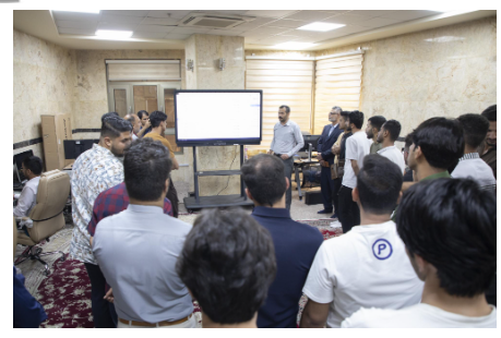
جامعة الكفيل تعقد ورشة عمل عن إجراءات عمل اللجان الامتحانيّة
|