آخر المواضيع المضافة

 النبات
الحيوان
الأحياء المجهرية
علم الأمراض
التقانة الإحيائية
التقنية الحيوية المكروبية
التقنية الحياتية النانوية
علم الأجنة
الأحياء الجزيئي
علم وظائف الأعضاء
الغدد
المضادات الحيوية
النبات
الحيوان
الأحياء المجهرية
علم الأمراض
التقانة الإحيائية
التقنية الحيوية المكروبية
التقنية الحياتية النانوية
علم الأجنة
الأحياء الجزيئي
علم وظائف الأعضاء
الغدد
المضادات الحيوية| Chondral Osteogenesis-Finger |


|
|
Read More
Date: 26-7-2016
Date: 26-7-2016
Date: 28-12-2020
|
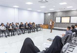
Chondral Osteogenesis-Finger
Longitudinal section through the index finger of a 6-month-old human embryo, with the base, middle and end of the limb to illustrate chondral osteogenesis (chondral, endochondral ossification), which begins in the dia-physis . Note the hyaline cartilage epiphyses. Chondral ossification is also referred to as indirect osteogenesis.
Stain: hematoxylin-eosin; magnification: × 18
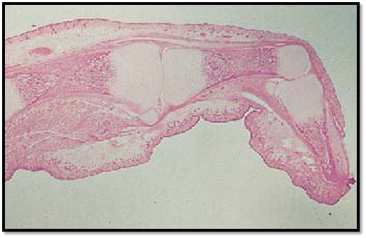
Chondral Osteogenesis-Finger
In contrast to the membranous, direct osteogenesis (ossification), the chondral osteogenesis requires an existing scaffold of hyaline cartilage lamellae ( cartilage model of ossification). While the cartilage is degrade d, it is replaced by bone tissue (substitute bone; indirect osteogenesis). Dependent on the lo-cation of their synthesis, there are perichondral and endochondral bones. The figure shows a longitudinal section through the middle phalanx of a finger. It presents the following details: the diaphysis 1 is in the center. Both the distal (left) and the proximal (right) epiphyses 2 still consist of hyaline cartilage (stained blue-violet). In the light zone between the two epiphyses, the diaphysis 1 , endochondral ossification has already started. The hyaline cartilage cells have changed into large spherical cells, and at the same time, calcium salts are deposited in the intercellular space. These areas can be recognized as small, branched, gray-blue lamellae. Osteoblasts reside in the connective tissue layer called perichondrium. They have built by membranous osteogenesis a sleeve of perichondral bone in the area of the dia-physis (here stained bright re d) 3 . The perichondrium is a form of connective tissue. The perichondral bone is a protective encasement for the bony structures in the area of the diaphysis. The two epiphyses are not yet affected by ossification.
1 Diaphysis
2 Epiphysis
3 Perichondral bone
4 Forming muscle cells
Stain: hematoxylin-eosin; magnification: × 15
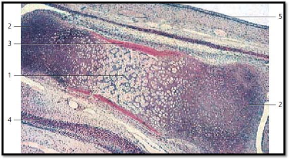
Chondral Osteogenesis—Finger
The perichondral bony sleeve 1 has become thicker by appositional growth . At the same time, it has expanded in the direction of the two epiphyses 2 . The cartilage cells in the diaphysis 3 have died, and the cartilage has disintegrated. In its place there is now vascular connective tissue, which has in-vaded from the outside. The tissue is mesenchymal in nature and is called primary bone marrow. Concurrently, small bony lamellae are created by chondral osteogenesis. In the direction of the epiphysis are rows of voluminous cartilage cells (column cartilage ), which stand out against the dormant cartilage tissue at the end of the joint .
1 Perichondral bone 3 Diaphysis
2 Epiphyseal cartilage 4 Periosteum
Stain: hematoxylin-eosin; magnification: × 12
References
Kuehnel, W.(2003). Color Atlas of Cytology, Histology, and Microscopic Anatomy. 4th edition . Institute of Anatomy Universitätzu Luebeck Luebeck, Germany . Thieme Stuttgart · New York .


|
|
|
|
دراسة يابانية لتقليل مخاطر أمراض المواليد منخفضي الوزن
|
|
|
|
|
|
|
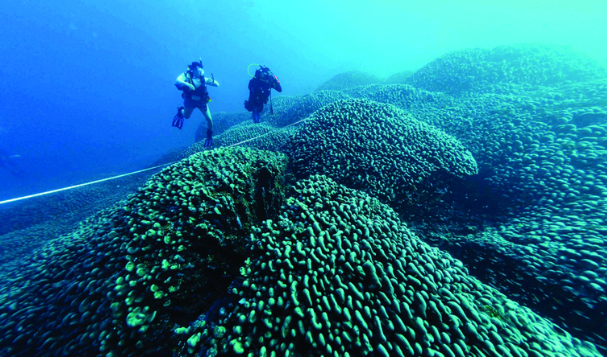
اكتشاف أكبر مرجان في العالم قبالة سواحل جزر سليمان
|
|
|
|
|
|
|
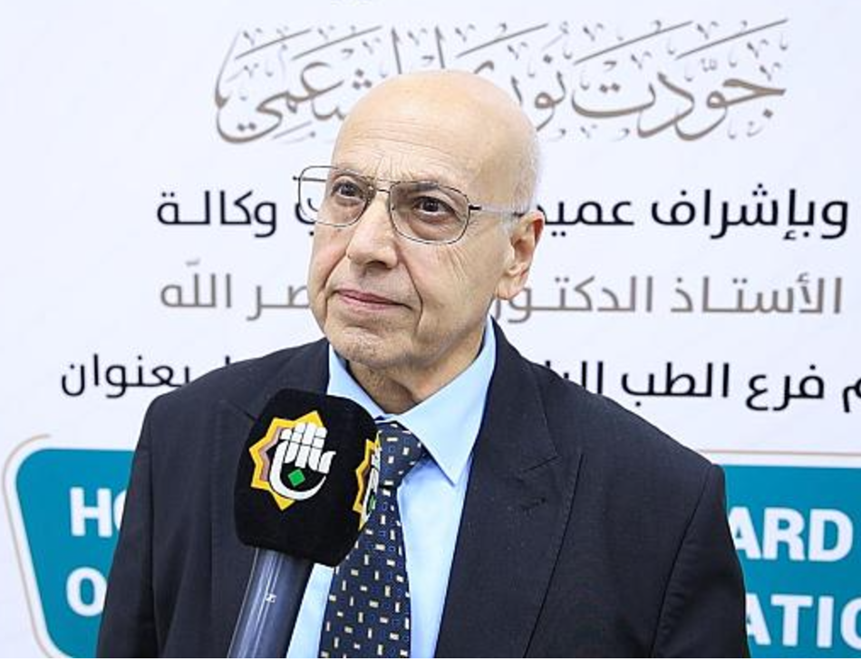
اتحاد كليات الطب الملكية البريطانية يشيد بالمستوى العلمي لطلبة جامعة العميد وبيئتها التعليمية
|
|
|