آخر المواضيع المضافة

 النبات
الحيوان
الأحياء المجهرية
علم الأمراض
التقانة الإحيائية
التقنية الحيوية المكروبية
التقنية الحياتية النانوية
علم الأجنة
الأحياء الجزيئي
علم وظائف الأعضاء
الغدد
المضادات الحيوية
النبات
الحيوان
الأحياء المجهرية
علم الأمراض
التقانة الإحيائية
التقنية الحيوية المكروبية
التقنية الحياتية النانوية
علم الأجنة
الأحياء الجزيئي
علم وظائف الأعضاء
الغدد
المضادات الحيوية| Eyelids-Palpebrae |


|
|
Read More
Date: 14-8-2016
Date: 1-8-2016
Date: 9-1-2017
|
Eyelids-Palpebrae
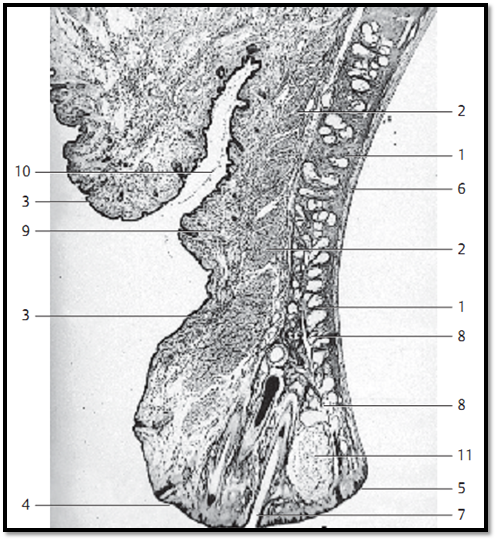
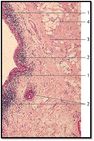
Eyelids are skin folds, which can be actively moved. They consist of a tough connective tissue skeleton 1 (tarsus superior and tarsus inferior ). Toward the outside, it is covered by the musculus orbicularis oculi (pars palpebralis) 2 . The surface covering of the eyelid is a multilayered keratinizing squamous epithelium with only a few velum hairs. The outer lid is about 2 mm wide and consists of a dull anterior 4 and a sharp-edge d posterior palpebral limb 5 . This tissue continues in the multilayered nonkeratinizing squamous epithelium of the palpebral part of the conjunctiva (conjunctiva tarsi) 6 . A multi-layered columnar epithelium with goblet cells is only found beyond the level of the fornix of the conjunctiva. Long cilia (eyelashes ) 7 protrude from the anterior rim of the lid. They are rooted in the lid plate. The sebaceous glands ( Zeis glands), apocrine scent glands and the sweat glands of the cilia ( Moll glands) end in the hair follicle of the eyelashes. The right side of the figure shows numerous tarsal holocrine sebaceous glands (Meibomian glands) 8 with long secretor y ducts that end on the anterior edge of the lid ( posterior limbus ). The tight tarsal fiber meshwork envelops the lobed glands. Smooth muscle cells run both before and behind the Meibomian glands at the rim of the lid. The palpebral part of the musculus orbicularis oculi 2 is located in front of the tarsus. The subcutaneous tissue of the lid consists of loosely structured, cell-rich connective tissue 9 , which is usually free of adipose tissue. The epithelium on the anterior eyelid is thin. The skin of the lids contains many melanocytes. This explains the dark pigmentation.
1 Tarsus
2 Palpebral part of the orbicularis oculi muscle
3 Skin
4 Anterior palpebral limb
5 Posterior palpebral limb
6 Palpebral part of the conjunctiva
7 Eyelashes
8 Tar sal glands, Meibomian glands
9 Loosely structured subcutaneous connective tissue
10 Deep skin fold of the upper lid
11 Chalazion (infection of the Meibomian gland)
Stain: hematoxylin-eosin; magnifying glass
Eyelids—Palpebrae
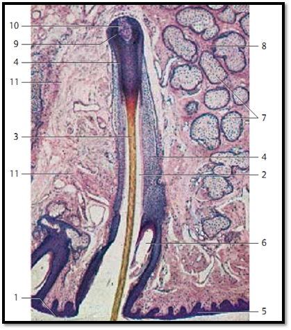
Detail magnification of an upper eyelid, with the rim of the lid and eyelashes. The following structures are shown:
1 Rim (edge) of the lid
2 Hair funnel
3 Hair shaft, scapus pili of the eyelash
4 Outer root sheath
5 Rim of the eyelid, multilayered keratinizing squamous epithelium
6 The terminal portions of a Meibomian gland end in the hair follicle clearance
8 Tarsus superior
9 Hair bulb
10 Hair papilla
11 Subcutaneous tissue of the lids
Stain: alum hematoxylin-eosin; magnification: × 10
Eyelids—Palpebrae
Detail magnification of a sagittal section through the eyelid of an adult
human. The skin on the outside of the lids is very thin. There is usually no adipose tissue 1 . It is malleable and can move laterally. Keratinization of the multilayered squamous epithelium of the epidermis is marginal. Dermis and subcutaneous connective tissue are thin. The dermis of the skin on the lid contains branched chromatophores . The palpebral part of the striated musculus orbicularis oculi 3 is shown in the lower right corner of the microphotograph. Part of the sebaceous follicle of a holocrine Meibomian gland 4 is visible in the upper right of the figure 4 .
1 Skin of the eyelid
2 Subcutaneous tissue
3 Palpebral part of the orbicularis oculi muscle
4 Meibomian gland
Stain: alum hematoxylin-eosin; magnification: × 20
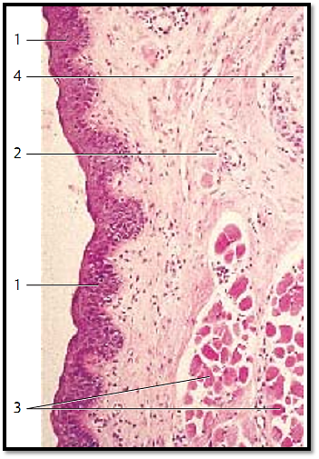
Eyelids—Palpebrae
The inner face of the eyelids is lined by the moist mucous membrane of the palpebral part of the tunica conjunctiva, which is part of the conjunctival sac. The keratinizing squamous epithelium of the skin of the lid continues in the nonkeratinizing multilayered squamous epithelium of the palpebral part of the conjunctiva at the posterior palpebral limb. It is only at the fornix conjunctivae that a multilayered columnar epithelium is found. The figure shows a sagittal section of the eyelid close to the fornix conjunctivae with conjunctival epithelium 1 and an underlying accumulation of lymphocytes 2 . The tunica propria 3 contains adipocytes 4 .
1 Conjunctival epithelium
2 Accumulation of lymphocytes
3 Lamina propria
4 Adipocytes
Stain: alum hematoxylin-eosin; magnification: × 20
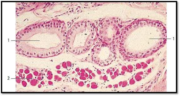
Eyelids—Palpebrae
Detail magnification of a sagittal section through the eyelid close to the rim of the lid. It shows the apocrine glands of Moll ( ciliary glands) 1 . They are located in the vicinity of the roots of the eyelashes and end at the rim of the lid or in the hair follicles. The glands of Moll are classified as sweat glands. There are striate d muscle fiber bundles of the musculus orbicularis oculi 2 close to the glands. They can pull the rim of the lid to the eyeball.
1 Glands of Moll, apocrine sweat glands
2 Orbicularis oculi muscle
Stain: alum hematoxylin-eosin; magnification: × 20
References
Kuehnel, W.(2003). Color Atlas of Cytology, Histology, and Microscopic Anatomy. 4th edition . Institute of Anatomy Universitätzu Luebeck Luebeck, Germany . Thieme Stuttgart · New York .


|
|
|
|
لخفض ضغط الدم.. دراسة تحدد "تمارين مهمة"
|
|
|
|
|
|
|
طال انتظارها.. ميزة جديدة من "واتساب" تعزز الخصوصية
|
|
|
|
|
|
|
مشاتل الكفيل تزيّن مجمّع أبي الفضل العبّاس (عليه السلام) بالورد استعدادًا لحفل التخرج المركزي
|
|
|