

 النبات
 الحيوان
 الأحياء المجهرية
 علم الأمراض
 التقانة الإحيائية
 التقنية الحيوية المكروبية
 التقنية الحياتية النانوية
 علم الأجنة
 الأحياء الجزيئي
 علم وظائف الأعضاء
 الغدد
الغدد
 المضادات الحيوية
|
Read More
Date: 17-1-2017
Date: 31-7-2016
Date: 3-8-2016
|
Olfactory Epithelium-Olfactory Region
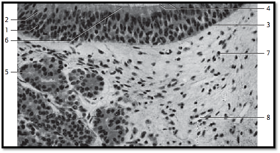
The epithelium of the olfactory mucous lamina is multilayered and contains specific sensory cells 1 , support cells 2 and basal cells 3 . The cones of the bi-polar olfactory cells 4 are visible in some areas of the epithelial surface. Olfactory glands 5 are present in the vascularize d innervated lamina propria. The serous glands consist of winding tubes. They release mucus to dissolve and remove olfactory materials.
1 Sensor y cells
2 Support cells
3 Basal cells
4 Conical olfactory cells
5 Olfactory glands
6 Glimpse of junctional complexes (terminal bars)
7 Plasma cell
8 Capillary
Stain: alum hematoxylin-eosin; magnification: × 100

Olfactory Epithelium—Olfactory Region
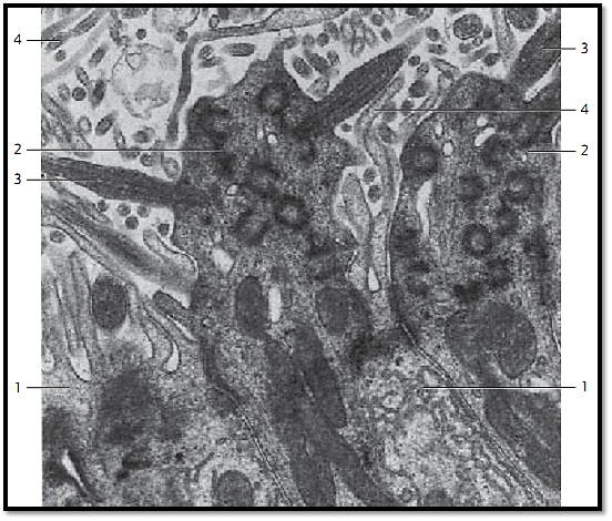
The multilayered columnar epithelium of the olfactory mucous lamina consists of basal cells, support cells and sensory olfactory cells . The nuclei of these cells occupy different levels. The support cells are the most numerous cells. Basal, support and sensor y cells originate on the basal lamina, forming a Pseudostratified epithelium. This figure shows the apical regions of two support cells and two sensor y cells. The support cells are usually wider at their apical surfaces than at their bases. Long microvilli 4 protrude from the free surfaces of support cells. They contain many organelles, a large Golgi apparatus, extended smooth ER and secretor y granules. The olfactory sensory cells are bipolar neurons. Their apex is wider than their basal part. Their olfactory bulbs extend over the epithelial surface. Six to eight long cilia 3 protrude from the olfactory cells, which contain the olfactory receptors. The base of each cilia shows the typica l9×2+2 microtubule organization. The microtubule continues in a thin nontubular process. The cilia protrude into a mucous film. Olfactory and support cells are connected via a network of terminal bars (tight junctions). Note the abundance of mitochondria in the sensory cells. The basal processes of the sensor y cells are axons, which run to the base of the epithelium and traverse the basal membrane. Apposed plasmalemma Schwann cell processes envelop the axons only after they have traverse d the basal lamina. The olfactory bulbs with their cilia make up the apical portion of the dendritic processes of these sensory cells.
1 Support cells with microvilli
2 Olfactory bulb with cilia
3 Kinocilia
4 Microvilli
Electron microscopy; magnification: × 24 000

Olfactory Epithelium—Olfactory Region
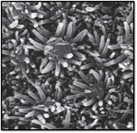
View of an area of the olfactory mucous lamina. The olfactory bulbs extend over the epithelial surface. Long cilia protrude from the olfactory bulbs. The sensory cells are interspersed with support cells with micro-villi. The olfactory bulbs are about 4 μm high.
Scanning electron microscopy; magnification: × 17 800

References
Kuehnel, W.(2003). Color Atlas of Cytology, Histology, and Microscopic Anatomy. 4th edition . Institute of Anatomy Universitätzu Luebeck Luebeck, Germany . Thieme Stuttgart · New York .


|
|
|
|
علامات بسيطة في جسدك قد تنذر بمرض "قاتل"
|
|
|
|
|
|
|
أول صور ثلاثية الأبعاد للغدة الزعترية البشرية
|
|
|
|
|
|
|
قسم الشؤون الفكرية والثقافية يجري اختبارات مسابقة حفظ دعاء أهل الثغور
|
|
|