آخر المواضيع المضافة

 النبات
الحيوان
الأحياء المجهرية
علم الأمراض
التقانة الإحيائية
التقنية الحيوية المكروبية
التقنية الحياتية النانوية
علم الأجنة
الأحياء الجزيئي
علم وظائف الأعضاء
الغدد
المضادات الحيوية
النبات
الحيوان
الأحياء المجهرية
علم الأمراض
التقانة الإحيائية
التقنية الحيوية المكروبية
التقنية الحياتية النانوية
علم الأجنة
الأحياء الجزيئي
علم وظائف الأعضاء
الغدد
المضادات الحيوية| Microvilli-Microplicae |


|
|
Read More
Date: 9-1-2017
Date: 31-7-2016
Date: 8-1-2017
|
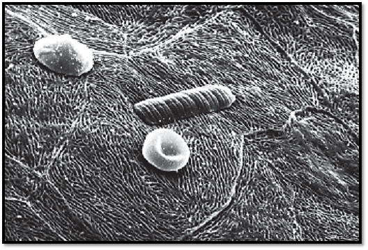
Microvilli-Microplicae
The cell surface can also be enlarged by raised, leaf-like formations of the plasmalemma—i.e., by outward extension in the form of folds. The figure shows flat epithelial cells from the canine tongue with a dense pattern of microplicae —i.e., raise d folds of the plasma membrane. Such microplicae folds are commonly observe d in a multitude of patterns. Microplicae also exist at the bottom face of flat epithelial cells, and therefore may play a role in intercellular adhesion. The more pronounced “edges” in this figure correspond to the cell borders. In transmission electron microscopy, vertical cuts show microplicae of ten as short, stump-shape d microvilli. Located on top of the epithelial cells are two erythrocytes and a rod-shaped form, which can be categorize d as an oral cavity saprophyte.
Scanning electron microscopy; magnification: × 3600
References
Kuehnel, W.(2003). Color Atlas of Cytology, Histology, and Microscopic Anatomy. 4th edition . Institute of Anatomy Universitätzu Luebeck Luebeck, Germany . Thieme Stuttgar t · New York .


|
|
|
|
الآثار الجانبية لأدوية تستخدم في علاج "ألزهايمر" تثير الجدل
|
|
|
|
|
|
|
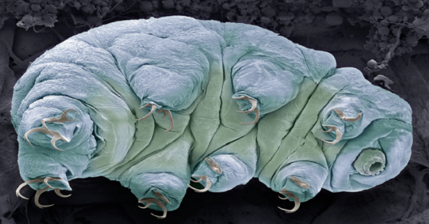
اكتشاف سر نجاة "مخلوقات أبدية" من انفجارات الإشعاع القاتلة
|
|
|
|
|
|
موكب أهالي كربلاء يهدي ممثل المرجعية العليا درعا تثمينا للمساهمات الفاعلة والمساندة لإنجاح الفعاليات التي يقيمها خلال المناسبات الدينية
|
|
|
|
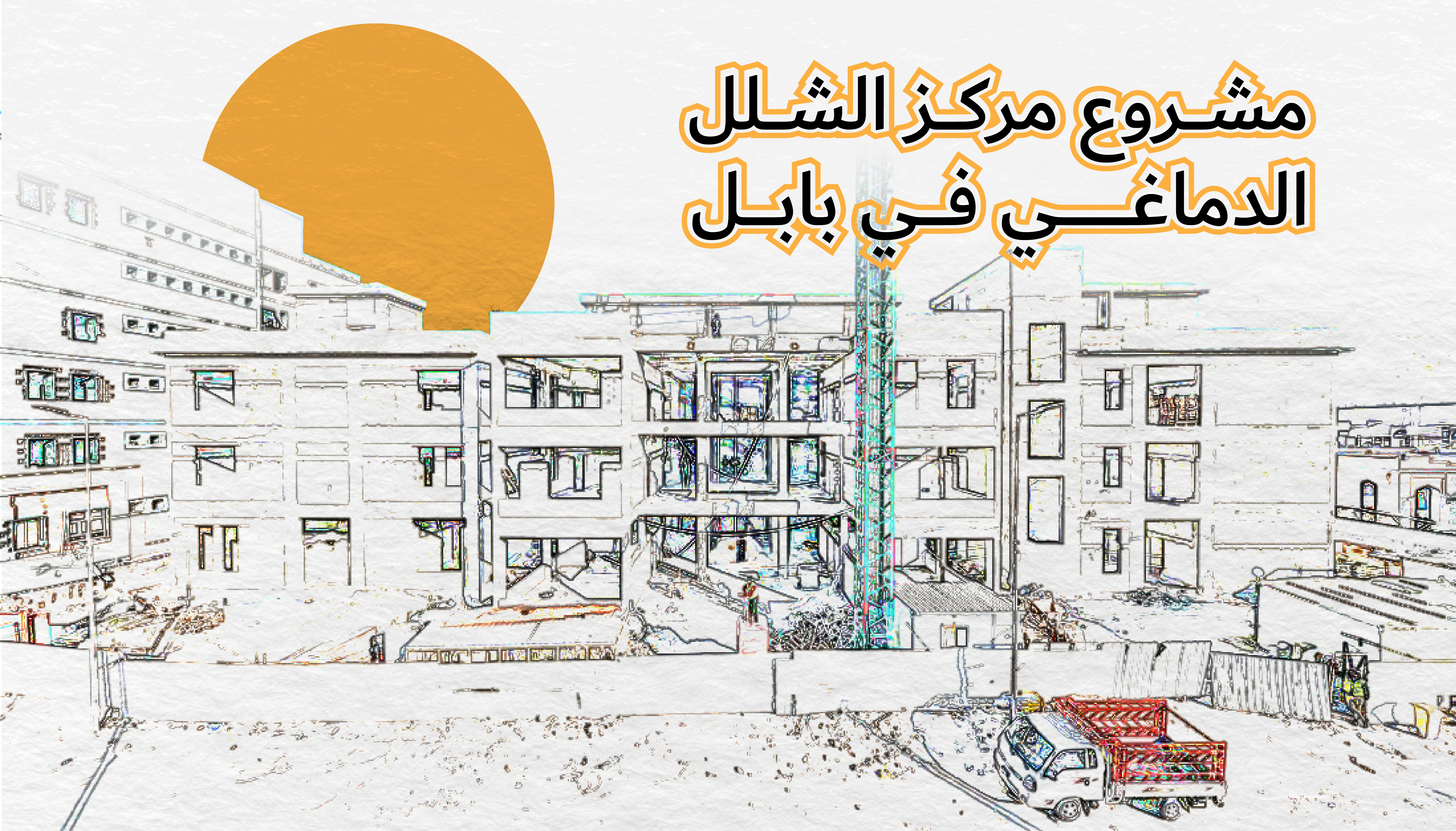
مراحل متقدمة من الإنجاز يشهدها مشروع مركز الشلل الدماغي في بابل
|
|
|
|
الأمين العام للعتبة الحسينية المقدسة: يجب الاهتمام بالباحثين عن العمل ومنحهم الفرص المناسبة عبر الاهتمام بقدراتهم ومؤهلاتهم وإبداعاتهم
|
|
|
|
يمتد على مساحة (500) دونم ويستهدف توليد الطاقة الكهربائية.. العتبة الحسينية تعلن عن الشروع بإنشاء مشروع معمل لتدوير النفايات في كربلاء
|