آخر المواضيع المضافة

 النبات
الحيوان
الأحياء المجهرية
علم الأمراض
التقانة الإحيائية
التقنية الحيوية المكروبية
التقنية الحياتية النانوية
علم الأجنة
الأحياء الجزيئي
علم وظائف الأعضاء
الغدد
المضادات الحيوية
النبات
الحيوان
الأحياء المجهرية
علم الأمراض
التقانة الإحيائية
التقنية الحيوية المكروبية
التقنية الحياتية النانوية
علم الأجنة
الأحياء الجزيئي
علم وظائف الأعضاء
الغدد
المضادات الحيوية| Cell death |


|
|
Read More
Date: 2025-03-11
Date: 26-2-2016
Date: 2025-01-13
|
Cell death
Cells can die via one of the following two ways:
1. Necrosis
2. Apoptosis
1. Necrosis
In necrosis, excess fluid enters the cell, swells it, & ruptures its membrane which kills it. After the cell has died, intracellular degradative reactions occur within a living organism. Necrosis does not occur in dead organisms. In dead organisms, autolysis & heterolysis take place.
Necrosis occurs by the following mechanisms:
A. Hypoxia
B. Free radical-induced cell injury
C. Cell membrane damage
D. Increased intracellular calcium level
A. Hypoxia
Hypoxia is decreased oxygen supply to tissues. It can be caused by:
1. Ischemia
Ischemia is decreased blood flow to or from an organ. Ischemia can be caused by obstruction of arterial blood flow – the most common cause, or by decreased perfusion of tissues by oxygen-carrying blood as occurs in cardiac failure, hypotension, & shock.
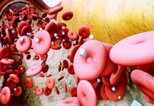
2. Anemia
Anemia is a reduction in the number of oxygen-carrying red blood cells.
3. Carbon monoxide poisoning
CO decreases the oxygen-capacity of red blood cells by chemical alteration of hemoglobin.
4. Poor oxygenation of blood due to pulmonary disease.
The cell injury that results following hypoxia can be divided into early & late stages:
1. Early (reversible) stages of hypoxic cell injury At this stage, hypoxia results in decreased oxidative phosphorylation & ATP synthesis. Decreased ATP leads to:
a. Failure of the cell membrane Na – K pump, which leads to increased intracellular Na & water, which cause cellular & organelle swelling. Cellular swelling (hydropic change) is characterized by the presence of large vacuoles in the cytoplasm. The endoplasmic reticulum also swells. The mitochondria show a low amplitude swelling. All of the above changes are reversible if the hypoxia is corrected.
b. Disaggregation of ribosomes & failure of protein synthesis.
2. Late (irreversible) stages of hypoxic cell injury.
This is caused by severe or prolonged injury. It is caused by massive calcium influx & very low pH, which lead to activation of enzymes, which damage the cell membrane& organelle membranes. Irreversible damage to the mitochondria, cell membranes, & the nucleus mark the point of no return for the cell, that is after this stage, the cell is destined to die.
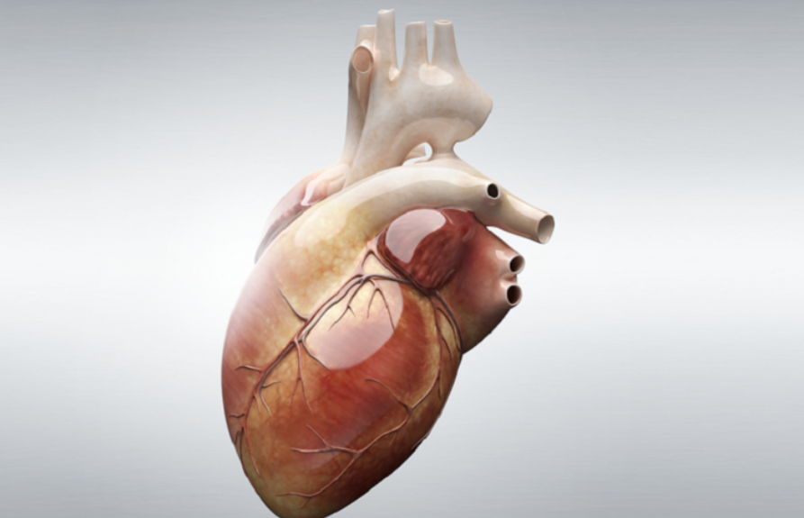
Release of aspartate aminotransferase (AST), creatine phosphokinase(CPK), & lactate dehydrogenase (LDH) into the blood is an important indicator of irreversible injury to heart muscle following myocardial infarction.
B. Free radical-induced injury
Free radical is any molecule with a single unpaired electron in the outer orbital. Examples include superoxide & the hydroxyl radicals. Free radicals are formed by normal metabolism, oxygen toxicity, ionizing radiation, & drugs & chemicals, & reperfusion injury. They are degraded by spontaneous decay, intracellular enzymes such as glutathione peroxidase, catalase, or superoxide dismutase, & endogenous substances such as ceruloplasmin or transferrin. When the production of free radicals exceeds their degradation, the excess free radicals cause membrane pump damage, ATP depletion, & DNA damage. These can cause cell injury & cell death.
C. Cell membrane damage
Direct cell membrane damage as in extremes of temprature, toxins, or viruses, or indirect cell membrane damage as in the case of hypoxia can lead to cell death by disrupting the homeostasis of the cell.
D. Increased intracellular calcium level
Increased intracellular calcium level is a common pathway via which different causes of cell injury operate. For example, the cell membrane damage leads to increased intracellular calcium level. The increased cytosolic calcium, in turn, activates enzymes in the presence of low pH. The activated enzymes will degrade the cellular organelles.
Types of necrosis
The types of necrosis include:
1. Coagulative necrosis
2. Liquefactive necrosis
3. Fat necrosis
4. Caseous necrosis
5. Gangrenous necrosis
1. Coagulative necrosis
Cogulative necrosis most often results from sudden interruption of blood supply to an organ, especially to the heart. It is, in early stages, characterized by general preservation of tissue architecture. It is marked by the following nuclear changes: Pyknosis (which is chromatin clumping & shrinking with increased basophilia), karyorrhexis (fragmentation of chromatin), & karyolysis (fading of the chromatin material).
2. Liquefactive necrosis
Liquefactive necrosis is characterized by digestion of tissue. It shows softening & liquefaction of tissue. It characteristically results from ischemic injury to the CNS. It also occurs in suppurative infections characterized by formation of pus.
3. Fat necrosis
Fat necrosis can be caused by trauma to tissue with high fat content, such as the breast or it can also be caused by acute hemorrhagic pancreatitis in which pancreatic enzymes diffuse into the inflamed pancreatic tissue & digest it. The fatty acids released from the digestion form calcium salts (soap formation or dystrophic calcification). In addition, the elastase enzyme digests the blood vessels & cause the hemorrhage inside the pancreas, hence the name hemorrhagic pancreatitis.
4. Caseous necrosis
Caseous necrosis has a cheese-like (caseous, white) appearance to the naked eye. And it appears as an amorphous eosinophilic material on microscopic examination. Caseous necrosis is typical of tuberculosis.
5. Gangrenous necrosis
This is due to vascular occlusion & most often affects the lower extremities & the bowel. It is called wet gangrene if it is complicated by bacterial infection which leads to superimposed liquefactive necrosis. Whereas it is called dry gangrene if there is only coagulative necrosis without liquefactive necrosis.
Necrosis can be followed by release of intracellular enzymes into the blood, inflammation or dystrophic calcification.
2. Apoptosis
Apoptosis is the death of single cells within clusters of other cells. (Note that necrosis causes the death of clusters of cells.) In apoptosis, the cell shows shrinkage & increased acidophilic staining of the cell. This is followed by fragmentation of the cells. These fragments are called apoptotic bodies. Apoptosis usually occurs as a physiologic process for removal of cells during embryogenesis, menstruation, etc… It can also be seen in pathological conditions caused by mild injurious agents.
Apoptosis is not followed by inflammation or calcification. The above mentioned features distinguish apoptosis from necrosis.
References
Bezabeh ,M. ; Tesfaye,A.; Ergicho, B.; Erke, M.; Mengistu, S. and Bedane,A.; Desta, A.(2004). General Pathology. Jimma University, Gondar University Haramaya University, Dedub University.


|
|
|
|
دخلت غرفة فنسيت ماذا تريد من داخلها.. خبير يفسر الحالة
|
|
|
|
|
|
|
ثورة طبية.. ابتكار أصغر جهاز لتنظيم ضربات القلب في العالم
|
|
|
|
|
|
|
سماحة السيد الصافي يؤكد ضرورة تعريف المجتمعات بأهمية مبادئ أهل البيت (عليهم السلام) في إيجاد حلول للمشاكل الاجتماعية
|
|
|