

 النبات
 الحيوان
 الأحياء المجهرية
 علم الأمراض
 التقانة الإحيائية
 التقنية الحيوية المكروبية
 التقنية الحياتية النانوية
 علم الأجنة
 الأحياء الجزيئي
 علم وظائف الأعضاء
 الغدد
الغدد
 المضادات الحيوية
|
Read More
Date: 16-12-2015
Date: 8-5-2016
Date: 25-2-2021
|
Carboxyl Proteinase
The carboxyl proteinases (E.C. 3.4.23) are also known as aspartyl proteinases or acid proteinases, so called because the mechanism by which they catalyze the hydrolysis of peptide bonds in proteins involves the participation of two carboxyl groups, usually provided by the side chains of two aspartic acid residues (1). The prototypical member of this large family of structurally-related enzymes is the gastric proteinase, pepsin, which is optimally active at acid pH. Other members include rennin (chymosin) from the fourth stomach of the calf; renin, a kidney enzyme found in blood plasma; cathepsin D, found in lysosomes; numerous fungal enzymes, such as penicillopepsin; and a number of retroviral proteinases from retroviruses, including the human immunodeficiency virus HIV proteinase that is the target of the proteinase inhibitors used in the treatment of acquired immune deficiency syndrome (AIDS). Pepsin and renin illustrate the range of specificity exhibited by proteinases in general and carboxyl proteinases in particular. Pepsin cleaves multiple bonds in virtually all proteins found in the diet, whereas renin cleaves a single bond in its only known biological substrate, angiotensinogen—the precursor of angiotensin.
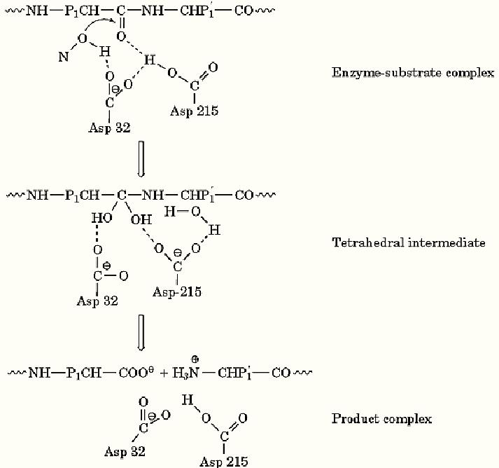
Carboxyl proteinases occur in all eukaryotic organisms and are important for protein processing and degradation. They are generally synthesized as inactive precursors that spontaneously activate under acidic conditions. Protein and peptide substrates bind to the active site of these enzymes, and one of the carboxylate groups (in the case of pepsin, it is from Asp32) activates a water molecule to attack the carboxyl group of the peptide bond that is to be cleaved (Figure 1). The other carboxyl group, which is protonated (in pepsin, it is Asp215), donates a proton to the peptide nitrogen atom to form an activated intermediate that dissociates into the products. It is believed that this general mechanism pertains to all of the carboxyl proteinases.

Figure 1. Schematic representation of the mechanism of action of carboxyl proteinases. The substrate (top line) binds to the active site of the enzyme [represented by the carboxyl groups of Asp32 and Asp215 (porcine pepsin numbering)] to form an enzyme-substrate complex. P1 and P′1 are the sidechains of the main specificity-determining amino acid residues that contribute the -CO- and -NH- groups to the peptide bond that will be hydrolyzed. The carboxylate group of Asp 32 promotes the attack of a water molecule on the carbonyl carbon of the peptide bond, to form a tetrahedral intermediate that is stabilized by transfer of a proton from Asp 215. The intermediate undergoes rearrangement to form a product complex, which subsequently dissociates, thereby regenerating the original enzyme.
It is interesting that in carboxyl proteinases such as pepsin and renin, both catalytic carboxyl groups are present in the same molecule. In the retroviral proteinase from HIV-1, which only contains 99 amino acids, two molecules form a dimer with a single active site containing one catalytic aspartic acid from each subunit. The larger mammalian and microbial carboxyl proteinases contain approximately 325 residues and appear to have homologous amino- and carboxyl-terminal domains that likely evolved from an early gene duplication and fusion event. Each domain contributes a catalytically active aspartic acid residue.
A characteristic feature of carboxyl proteinases is their susceptibility to inhibition by pepstatin, an acylated pentapeptide analogue produced by Streptomyces. Recently a number of carboxyl proteinase inhibitors have been found useful for the treatment of AIDS, particularly when used in conjunction with other viral enzyme inhibitors. Carboxyl proteinase inhibitors have also been proposed for the treatment of malaria. Two such proteinases are believed to be essential for degradation of hemoglobin in the red blood cell phase of the life cycle of the parasite.
References
1. D. R. Davies (1990) Annu. Rev. Biophys. Biophys. Chem. 19, 189–215.


|
|
|
|
علامات بسيطة في جسدك قد تنذر بمرض "قاتل"
|
|
|
|
|
|
|
أول صور ثلاثية الأبعاد للغدة الزعترية البشرية
|
|
|
|
|
|
|
مكتبة أمّ البنين النسويّة تصدر العدد 212 من مجلّة رياض الزهراء (عليها السلام)
|
|
|