

 النبات
 الحيوان
 الأحياء المجهرية
 علم الأمراض
 التقانة الإحيائية
 التقنية الحيوية المكروبية
 التقنية الحياتية النانوية
 علم الأجنة
 الأحياء الجزيئي
 علم وظائف الأعضاء
 الغدد
الغدد
 المضادات الحيوية
|
Read More
Date: 6-11-2015
Date: 1-11-2015
Date: 6-11-2015
|
T Cells
T-Cell Activation
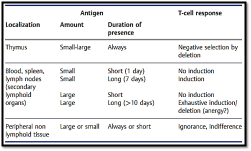
There are two classes of T cells; T helper cells (CD4+) and cytotoxic T cells (CD8+). Table 1 summarizes the reliance of T-cell responses on the dose, localization, and duration of presence of antigen. T-cell stimulation via the TCR, accessory molecules and adhesion molecules results in the activation
Table 1 Dependence of T-Cell Response on Antigen Localization, Amount and Duration of Presence

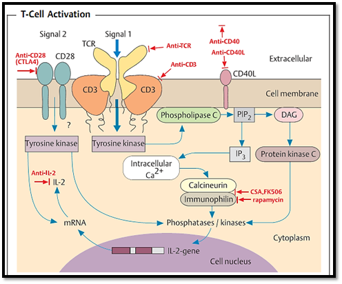
of various tyrosine kinases (Fig. 1) and mediates stringent and differential regulation of several signaling steps. T-cell induction and activation result from the activation of two signals. In addition to TCR activation (signal 1 = antigen), a costimulatory signal (signal 2) is usually required. Important costimulatory signals are delivered by the binding of B7 (B7.1 and B7.2) proteins (present on the APC or B cell) to ligands on the Tcells (CD28 protein, CTLA-4), or by CD40-CD40 ligand interactions. T-cell expansion is also enhanced by 1L-2.

Fig. 1 Regulation of T-cell activation is controlled by multiple signals, including costimulatory signals (Signal 2). Stimulation of the T cell via the T-cell receptor (TCR; Signal 1) activates a tyrosine kinase, which in turn activates phospholipase C (PLC). PLC splits phosphatidylinositol bisphosphate (PIP2) into inositol trispho- sphate (IP3) and diacyl glycerol (DAG). IP3 releases Ca2+ from intracellular depots, whilst DAG activates protein kinase C (PKC). Together, Ca2+ and PKC induce and activate the phosphoproteins required for IL-2 gene transcription within the cell nucleus. Stimulation of a T cell via the TCR alone results in production of only very small amounts of IL-2. Increased IL-2 production often requires additional signals (costimulation, e.g., via CD28). Costimulation via CD28 activates tyrosine kinases, which both sustain the transcription process and ensure post-transcriptional stabilization of IL-2 mRNA. Immunosuppressive substances (in red letters) include cytostatic drugs, anti-TCR, anti-CD3, anti-CD28 (CTLA4), anti-CD40, cyclosporine A and FK506 (which interferes with immunophilin-calcineurin binding, thus reducing IL-2 production), and rapamycin (which binds to, and blocks, immunophilin and hardly reduces IL-2 at all). Anti-interleukins (especially anti-IL-2, or a combination of anti-IL-2 receptor and anti-IL-15) block T-cell proliferation.
T-Cell Activation by Superantigens
In association with MHC class 11 molecules, a number of bacterial and possibly viral products can efficiently stimulate a large repertoire of CD4+ T cells at one time. This is often mediated by the binding of the bacterial or viral product to the constant segment of certain VP chains (and possibly Va chains) with a low level of specificity. Superantigens are categorized as either exogenous or endogenous. Exogenous superantigens mainly include bacterial toxins (staphylococcus enterotoxin types A-E [SEA, SEB, etc.]), toxic shock syndrome toxin (TSST), toxins from Streptococcus pyogenes, and certain retroviruses. Endogenous superantigens are derived from components of certain retroviruses found in mice, and which display superantigen-like behavior (e.g., murine mammary tumor virus, MMTV). The function of superantigens during T cell activation can be compared to the effect of bacterial lipopolysaccharides on B cells, in that LPS-induced B cell activation is also polyclonal (although it functions by way of the LPS receptors instead of the 1g receptors.
References
Zinkernagel, R. M. (2005). Medical Microbiology. © Thieme.


|
|
|
|
دراسة يابانية لتقليل مخاطر أمراض المواليد منخفضي الوزن
|
|
|
|
|
|
|
اكتشاف أكبر مرجان في العالم قبالة سواحل جزر سليمان
|
|
|
|
|
|
|
اتحاد كليات الطب الملكية البريطانية يشيد بالمستوى العلمي لطلبة جامعة العميد وبيئتها التعليمية
|
|
|