آخر المواضيع المضافة

 النبات
الحيوان
الأحياء المجهرية
علم الأمراض
التقانة الإحيائية
التقنية الحيوية المكروبية
التقنية الحياتية النانوية
علم الأجنة
الأحياء الجزيئي
علم وظائف الأعضاء
الغدد
المضادات الحيوية
النبات
الحيوان
الأحياء المجهرية
علم الأمراض
التقانة الإحيائية
التقنية الحيوية المكروبية
التقنية الحياتية النانوية
علم الأجنة
الأحياء الجزيئي
علم وظائف الأعضاء
الغدد
المضادات الحيوية| Microvilli-Uterus |


|
|
Read More
Date: 25-7-2016
Date: 14-8-2016
Date: 3-8-2016
|
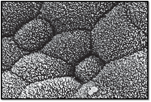
Microvilli-Uterus
Many epithelial cells form processes, name d microvilli , at their free surfaces. These processes are finger-shaped; they are about 100 nm thick and of various lengths. The structure of microvilli is static and without any particular organization. However, protrusions from the plasmalemma increase the cell surface area. Scanning electron microscopy provides a method of examining the outer elements of the tissue surface in detail. This figure offers a view of the surface of the uterine cavum epithelium. The cells are either polygonal or round and covered with short stub-like microvilli. The image is that of small patches of lawn. The dark lines between the cells are cell borders. In the lower right part of the figure are two erythrocytes.
Scanning electron microscopy; magnification: × 3000
References
Kuehnel, W.(2003). Color Atlas of Cytology, Histology, and Microscopic Anatomy. 4th edition . Institute of Anatomy Universitätzu Luebeck Luebeck, Germany . Thieme Stuttgar t · New York .


|
|
|
|
التوتر والسرطان.. علماء يحذرون من "صلة خطيرة"
|
|
|
|
|
|
|
مرآة السيارة: مدى دقة عكسها للصورة الصحيحة
|
|
|
|
|
|
|
نحو شراكة وطنية متكاملة.. الأمين العام للعتبة الحسينية يبحث مع وكيل وزارة الخارجية آفاق التعاون المؤسسي
|
|
|