
النبات

مواضيع عامة في علم النبات

الجذور - السيقان - الأوراق

النباتات الوعائية واللاوعائية

البذور (مغطاة البذور - عاريات البذور)

الطحالب

النباتات الطبية


الحيوان

مواضيع عامة في علم الحيوان

علم التشريح

التنوع الإحيائي

البايلوجيا الخلوية


الأحياء المجهرية

البكتيريا

الفطريات

الطفيليات

الفايروسات


علم الأمراض

الاورام

الامراض الوراثية

الامراض المناعية

الامراض المدارية

اضطرابات الدورة الدموية

مواضيع عامة في علم الامراض

الحشرات


التقانة الإحيائية

مواضيع عامة في التقانة الإحيائية


التقنية الحيوية المكروبية

التقنية الحيوية والميكروبات

الفعاليات الحيوية

وراثة الاحياء المجهرية

تصنيف الاحياء المجهرية

الاحياء المجهرية في الطبيعة

أيض الاجهاد

التقنية الحيوية والبيئة

التقنية الحيوية والطب

التقنية الحيوية والزراعة

التقنية الحيوية والصناعة

التقنية الحيوية والطاقة

البحار والطحالب الصغيرة

عزل البروتين

هندسة الجينات


التقنية الحياتية النانوية

مفاهيم التقنية الحيوية النانوية

التراكيب النانوية والمجاهر المستخدمة في رؤيتها

تصنيع وتخليق المواد النانوية

تطبيقات التقنية النانوية والحيوية النانوية

الرقائق والمتحسسات الحيوية

المصفوفات المجهرية وحاسوب الدنا

اللقاحات

البيئة والتلوث


علم الأجنة

اعضاء التكاثر وتشكل الاعراس

الاخصاب

التشطر

العصيبة وتشكل الجسيدات

تشكل اللواحق الجنينية

تكون المعيدة وظهور الطبقات الجنينية

مقدمة لعلم الاجنة


الأحياء الجزيئي

مواضيع عامة في الاحياء الجزيئي


علم وظائف الأعضاء


الغدد

مواضيع عامة في الغدد

الغدد الصم و هرموناتها

الجسم تحت السريري

الغدة النخامية

الغدة الكظرية

الغدة التناسلية

الغدة الدرقية والجار الدرقية

الغدة البنكرياسية

الغدة الصنوبرية

مواضيع عامة في علم وظائف الاعضاء

الخلية الحيوانية

الجهاز العصبي

أعضاء الحس

الجهاز العضلي

السوائل الجسمية

الجهاز الدوري والليمف

الجهاز التنفسي

الجهاز الهضمي

الجهاز البولي


المضادات الميكروبية

مواضيع عامة في المضادات الميكروبية

مضادات البكتيريا

مضادات الفطريات

مضادات الطفيليات

مضادات الفايروسات

علم الخلية

الوراثة

الأحياء العامة

المناعة

التحليلات المرضية

الكيمياء الحيوية

مواضيع متنوعة أخرى

الانزيمات
Edema
المؤلف:
Bezabeh ,M. ; Tesfaye,A.; Ergicho, B.; Erke, M.; Mengistu, S. ; Bedane,A. and Desta, A
المصدر:
General Pathology
الجزء والصفحة:
23-2-2016
2665
Edema
Definition: Edema is increased fluid in the interstitial tissue spaces or it is a fluid accumulation in the body cavities in excessive amount.
Depending on the site, fluid accumulation in body cavities can be variously designated as:
a) Hydrothorax – fluid accumulation in pleural cavity in a pathologic amount.
b) Hydropericardium – pathologic amount of fluid accumulated in the pericardial cavity.
c) Hydroperitoncum (ascites) – fluid accumulation in peritoneal cavity.
d) Ancsarca – is a severe & generalized edema of the body with profound
subcutaneous swelling.
Mechanism of edema formation:
Approximately 60% of the lean body weight is water, two-thirds of which is intracellular with
the remainder in the extracellular compartment.
The capillary endothelium acts as a semipermeable membrane and highly permeable to
water & to almost all solutes in plasma with an exception of proteins. Proteins in plasma
and interstial fluid are especially important in controlling plasma & interstitial fluid volume.
Normally, any outflow of fluid into the interstitium from the arteriolar end of the
microcirculation is nearly balanced by inflow at the venular end. Therefore, normally, there
is very little fluid in the interstitium.
Edema formation is determined by the following factors:
1) Hydrostatic pressure
2) Oncotic pressure
3) Vascular permeability
4) Lymphatic channels
5) Sodium and water retention
We will discuss each of the above sequentially.
1) Hydrostatic and oncotic pressures:
The passage of fluid across the wall of small blood vessels is determined by the balance between hydrostatic & oncotic pressures.
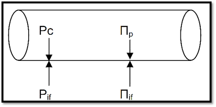
There are four primary forces that determine fluid movement across the capillary membrane. Each of them can be listed under the above two basic categories, the hydrostatic pressure & the oncotic pressure. These four primary forces are known as Starling forces & they are:
a. The capillary hydrostatic pressure (Pc)
This pressure tends to force fluid outward from the intravascular space through the capillary membrane to the interstitium.
b. The interstial fluid hydrostatic pressure (Pif)
This pressure tends to force fluid from the interstitial space to the intravascular space.
c. The plasma colloid osmotic (oncotic) pressure (Пp)
This pressure tends to cause osmosis of fluid inward through the capillary
membrane from the interstitium. The plasma oncotic pressure is caused by the presence of plasma proteins.
d. The interstial fluid colloid osmotic (oncotic) pressure (Пif) This pressure tends to cause osmosis of fluid outward through the capillary membrane to the interstitium.

Fig. 1 The forces that determine the movement of fluid across the capillary wall.
The net filtration pressure can be calculated as

In addition, some fluid is normally derained by the lymphatic channels. Usually, excess fluid will accumulate in the interstitium (i.e. edema is formed) when the capillary hydrostatic pressure is increased or when the plama oncotic pressure is decreased or when the lymphatic drainage is blocked.
Hence, basically, one can divide pathologic edema into two broad categories:
A. Edema due to decreased plasma oncotic pressure. The plasma oncotic pressure is decreased when the plasma proteins are decreased in various diseases such as:
1. Protein loosing glomerulopathies like nephroticsyndrome with leaky glomerulus.
2. Liver cirrhosis which leads to decreased protein synthesis by the damaged liver.
3. Malnutrition
4. Protein loosing enteropathy.
B. Edema resulting from increased capillary hydrostatic pressure as in the following diseases:
1. Deep venous thrombosis resulting in impaired venous return.
2. Pulmonary oedema
3. Cerebral oedema
4. Congestive heart failure
Clinical classification of edema:
One can also clinically classify edema into localized & generalized types.
A) Localized B) Generalized
1) Deep venous thrombosis 1) Nephrotic syndrome
2) Pulmonary edema 2) Liver cirrhosis
3) Brain edema 3) Malnutrition
4) Lymphatic edema 4) Heart failure
5) Renal failure
Next, we will elaborate on some of the above examples.
1. Localized edema
a. Edema of the brain:
- May be localized at the site of lesion e.g neoplasm, trauma.
- May be generalized in encephalitis, hypertensive crisis, & trauma
- Narrowed sulci & distended gyri.
- ↑ Edema → compression of medulla towards formen magnum → compression of vital centers lead to →- Hernation of the brain
↓
Patient dies
b. Pulmonary edema
- Usually occurs in left ventricular failure.
- May occur in adult respiratory distress syndrome (ARDS).
- lung ↑ 2.3x its weight.
2. Generalized edema (anasarca) occurs due to
a. Reduction of albumin due to excessive loss or reduced synthesis as is caused by:
1) Protein loosing glomerulopathies like nephrotic syndrome
2) Liver cirrhosis
3) Malnutrition
4) Protein-losing enteropathy
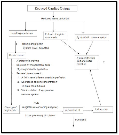
b. Increased volume of blood secondary to sodium retention caused by congestive heart failure:
Fig. 2. Mechanism of edema formation in congestive heart failure:

- From angiotensinogen
- α Globulin present in plasma
a. stimulate release of aldosterone
b. Causes vasoconstriction
c. degraded to angiotensin III which has similar functions
NB: If perfusion fails to improve, this cycle will operate continuously further exacerbating the edema resulting in anasarca.
This ends our discussion of the first two factors which determine edema formation. We will now go on to discuss the other factors.
2) Vascular permeability:
Increased vascular permeability usually occurs due to acute inflammation. In inflammation, chemical mediators are produced. Some of these mediators cause increased vascular permeability which leads to loss of fluid & high molecular weight albumin and globulin into the interstitium. Such edema (i.e. that caused by increased vascular permeability) is called inflammatory edema. Inflammatory edema differs from non-inflammatory edema by the following features
a) Inflammatory edema (exudate)
⇒ Due to inflammation-induced increased permeability and leakage of plasma proteins.
⇒ Forms an exudate [protein rich]
⇒ Specific gravity > 1.012
b) Non-inflammatory oedema (transudate)
⇒ A type of edema occurring in hemodynamic derangement (i.e. increased plasma hydrostatic pressure & decreased plasma oncotic pressure. See above)
⇒ Formed transudate [protein poor]
⇒ Specific gravity < 1.012
3) Lymphatic channels:
Also important is the lymphatic system which returns to the circulation the small amount of proteinaceous fluid that does leak from the blood into the interstial spaces. Therefore, obstruction of lymphatic channels due to various causes leads to the accumulation of the proteinaceous fluid normally drained by the lymphatic channels. Such kind of edema is called lymphatic edema.
Lymphatic edema occurs in the following conditions:
1) Parasitic infection. E.g filariasis which causes massive lymphatic and inguinal fibrosis
2) Lymphatic obstruction secondary to neoplastic infiltration. E.g. breast cancer 3) post surgical or post irradiation, i.e surgical resection of lymphatic channels or scarring after irradiation
4) Sodium and water retention:
Sodium & subsequently water retention occurs in various clinical conditions such as congetive heart failure (See Fig. 2, above) & renal failure. In these conditions, the retained sodium & water result in increased capillary hydrostatic pressure which leads to the edema seen in these diseases.
Morphology of edema
Microscopy
- Manifests only as subtle cell swelling. Clearing & separation of extracellular matrix.
References
Bezabeh ,M. ; Tesfaye,A.; Ergicho, B.; Erke, M.; Mengistu, S. and Bedane,A.; Desta, A.(2004). General Pathology. Jimma University, Gondar University Haramaya University, Dedub University.
 الاكثر قراءة في اضطرابات الدورة الدموية
 اخر الاخبار
اخبار العتبة العباسية المقدسة

الآخبار الصحية


 قسم الشؤون الفكرية يصدر كتاباً يوثق تاريخ السدانة في العتبة العباسية المقدسة
 "المهمة".. إصدار قصصي يوثّق القصص الفائزة في مسابقة فتوى الدفاع المقدسة للقصة القصيرة
 (نوافذ).. إصدار أدبي يوثق القصص الفائزة في مسابقة الإمام العسكري (عليه السلام)


