آخر المواضيع المضافة

 النبات
الحيوان
الأحياء المجهرية
علم الأمراض
التقانة الإحيائية
التقنية الحيوية المكروبية
التقنية الحياتية النانوية
علم الأجنة
الأحياء الجزيئي
علم وظائف الأعضاء
الغدد
المضادات الحيوية
النبات
الحيوان
الأحياء المجهرية
علم الأمراض
التقانة الإحيائية
التقنية الحيوية المكروبية
التقنية الحياتية النانوية
علم الأجنة
الأحياء الجزيئي
علم وظائف الأعضاء
الغدد
المضادات الحيوية| Imaging in Medicine |


|
|
Read More
Date: 26-10-2015
Date: 28-12-2020
Date: 9-10-2015
|
Imaging in Medicine
As recently as in the 1970s, the diagnosis of some diseases often required exploratory surgery, opening a body cavity to “have a look around” for visible disorders. The risks of infection, anesthesia, and imperfect healing weigh against exploratory surgery, but the diagnostic benefit may make the risk worth taking. In the last few decades, however, a variety of medical imaging techniques has made most exploratory surgery unnecessary and has greatly accelerated progress in medicine. Although the basic principles of some of these techniques have been known for much longer, they did not become clinically useful until computer technology had advanced enough to process data into clear images of the body, mostly since the 1970s.
Radiography
Radiography, use of X rays, is the oldest imaging technique. The term “X ray” can refer either to the type of radiation used or to the photographic image produced (the radiogram). X rays were discovered in 1885, and Marie Curie (1867-1934) trained military doctors in the use of X-ray machines in World War I. X rays are relatively simple and inexpensive to make, and they are commonly used in dentistry, mammography, chest examinations, and diagnosis of fractures. They are best used for dense structures such as bone, but hollow organs can be visualized by filling them with a radiopaque substance such as barium, given by swallow or enema to X ray the stomach or colon. Angiography is the X-ray visualization of blood vessels after injection with a radiopaque dye.
Sonography
Sonography, or ultrasound imaging, is the second oldest imaging method, and the second most widely used. An outgrowth of the sonar technology developed in World War II, it uses a handheld probe to “bombard” the body with ultrasound waves and a computer to analyze the reflected signal into an image. Sonography avoids the harmful effects of X rays and is commonly used to examine fetuses.
Computed Tomography (CT)
Formerly called a CAT scan, computed tomography (CT) is a more sophisticated use of X rays to produce more finely detailed images. Thepatient is moved through a machine that emits low-intensity X rays on one side and receives them with a detector on the other side. By imaging body slices as thin as a coin, CT scans show less overlap of organs than conventional X rays and thus produce sharper images. CT scans are useful for identifying tumors, aneurysms, cerebral hemorrhages, kidney stones, and other disorders.
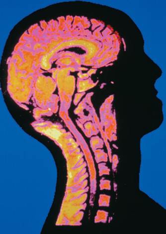
A magnetic resonance imaging (MRI) scan of a human brain, cervical spine, and spinal marrow.
Magnetic Resonance Imaging (MRI)
With magnetic resonance imaging (MRI), a cylindrical device surrounds the body with a magnetic field three thousand to sixty thousand times as strong as Earth’s. Hydrogen atoms align themselves with this field. The patient is then irradiated with radio waves. Hydrogen ions absorb this energy and align in a new direction. When the radio waves are turned off, they realign to the magnetic field and emit energy at rates that vary with the type of tissue. This emitted energy is received by a detector and analyzed by a computer into an image of the body’s interior. MRI can see through cranial and vertebral bone to visualize brain and spinal cord tissue in finer detail than CT.
Positron Emission Tomography (PET)
Positron emission tomography (PET) is used to visualize the metabolic state of a tissue. The patient receives an injection of radioactively labeled glucose, which emits charged particles called positrons. When a positron and electron meet, they annihilate each other and give off gamma rays that are picked up by a detector and analyzed by computer. The result is a color- coded image that shows which tissues were using the most glucose (that is, were most metabolically active) at the time. In cardiology, a PET scan can show the location and extent of dead heart tissue. In neuroscience, it can show which parts of the brain are active from moment to moment as a person engages in various sensory, motor, or intellectual tasks.
Functional MRI (fMRI)
A new variation of MRI, functional MRI (fMRI) detects the anaerobic activity of active neurons of the brain. It can pinpoint brain activity to within 1 or 2 millimeters, and is even more precise and useful than PET scans for studies of brain function. It also has the advantage of requiring no injections or radioactive isotopes, and it is much quicker than a PET scan. The PET and fMRI techniques not only have been valuable for clinical diagnosis but have added enormously to our knowledge of brain function, pinpointing abnormalities correlated with depression, schizophrenia, and attention deficit disorder. They have also provided images of the mind at work, so to speak, identifying areas involved in consciousness, memory, thought, musical perception, reading, motor control, and speech.
Radiology is the medical specialty that embraces all of these imaging techniques. Nuclear medicine is a branch of medicine that uses radioisotopes in the making of medical images, as in PET scans, and in the treatment of diseases such as cancer. Noninvasive techniques are those that require no break in the body surface whatsoever: conventional X rays; sonography; and CT, MRI, and fMRI scans. If a technique involves even such a slight invasion of the body as an injection or a barium swallow, it is considered an invasive procedure (angiography and PET scans, for example).
References
Brant, William E., and Clyde A. Helms, eds. Fundamentals of Diagnostic Radiology, 2nd ed. Philadelphia, PA: Lippincott, Williams and Wilkins, 1999.
National Institutes of Health, National Library of Medicine. Visible Human Project. <http://www.nlm.nih.gov/research/visible/visible_human.html>.
Weisslader, Ralph, MarkJ. Rieumont, and Jack Wittenberg. Primer of Diagnostic Imaging, 2nd ed. St. Louis, MO: Mosby, 1997.


|
|
|
|
دخلت غرفة فنسيت ماذا تريد من داخلها.. خبير يفسر الحالة
|
|
|
|
|
|
|
ثورة طبية.. ابتكار أصغر جهاز لتنظيم ضربات القلب في العالم
|
|
|
|
|
|
|
بالتعاون مع العتبتين المقدستين.. أهالي كربلاء يحيون ذكرى هدم قبور أئمة البقيع (عليهم السلام)
|
|
|