

 علم الكيمياء
 الكيمياء التحليلية
 الكيمياء الحياتية
 الكيمياء العضوية
 الكيمياء الفيزيائية
 الكيمياء اللاعضوية
 مواضيع اخرى في الكيمياء
 الكيمياء الصناعية
 |
Read More
Date: 9-3-2020
Date: 11-3-2020
Date: 22-2-2020
|
The crystals that form are frozen in liquid nitrogen and taken to the synchrotron which is a highly powered tunable x-ray source. They are mounted on a goniometer and hit with a beam of x-rays. Data is collected as the crystal is rotated through a series of angles. The angle depends on the symmetry of the crystal.
Figure 1: Top Left) This is a picture of a protein crystal mounted on a loop with respect to the UC Davis Structural Biology Lab; Bottom Right) This is a diffraction pattern created from the APS Kinase D63N Mutant of the above crystal with respect to the UC Davis Structural Biology Lab
Proteins are among the many biological molecules that are used for x-ray Crystallography studies. They are involved in many pathways in biology, often catalyzing reactions by increasing the reaction rate. Most scientists use x-ray Crystallography to solve the structures of protein and to determine functions of residues, interactions with substrates, and interactions with other proteins or nucleic acids. Proteins can be co - crystallized with these substrates, or they may be soaked into the crystal after crystallization.

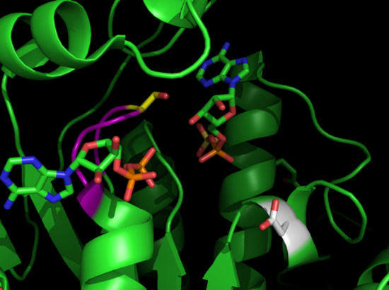
Figure 9: Top Left) This is the structure of APS Kinase co - crystallized with ligands ADP and APS created via pymol by an undergrad working in the Structural Biology lab at UC Davis; Bottom right) This is the mutant overlay of APS kinase. The teal is the wild - type and the lime green is the mutant. D63 (from the wild-type) is mutated to asparagine. Images created by pymol by an undergrad working in the Structural Biology lab at UC Davis.


|
|
|
|
التوتر والسرطان.. علماء يحذرون من "صلة خطيرة"
|
|
|
|
|
|
|
مرآة السيارة: مدى دقة عكسها للصورة الصحيحة
|
|
|
|
|
|
|
نحو شراكة وطنية متكاملة.. الأمين العام للعتبة الحسينية يبحث مع وكيل وزارة الخارجية آفاق التعاون المؤسسي
|
|
|