


النبات

مواضيع عامة في علم النبات
الجذور - السيقان - الأوراق
النباتات الوعائية واللاوعائية
البذور (مغطاة البذور - عاريات البذور)
الطحالب
النباتات الطبية
الحيوان
مواضيع عامة في علم الحيوان
علم التشريح
التنوع الإحيائي
البايلوجيا الخلوية
الأحياء المجهرية
البكتيريا
الفطريات
الطفيليات
الفايروسات
علم الأمراض
الاورام
الامراض الوراثية
الامراض المناعية
الامراض المدارية
اضطرابات الدورة الدموية
مواضيع عامة في علم الامراض
الحشرات
التقانة الإحيائية
مواضيع عامة في التقانة الإحيائية
التقنية الحيوية المكروبية
التقنية الحيوية والميكروبات
الفعاليات الحيوية
وراثة الاحياء المجهرية
تصنيف الاحياء المجهرية
الاحياء المجهرية في الطبيعة
أيض الاجهاد
التقنية الحيوية والبيئة
التقنية الحيوية والطب
التقنية الحيوية والزراعة
التقنية الحيوية والصناعة
التقنية الحيوية والطاقة
البحار والطحالب الصغيرة
عزل البروتين
هندسة الجينات
التقنية الحياتية النانوية
مفاهيم التقنية الحيوية النانوية
التراكيب النانوية والمجاهر المستخدمة في رؤيتها
تصنيع وتخليق المواد النانوية
تطبيقات التقنية النانوية والحيوية النانوية
الرقائق والمتحسسات الحيوية
المصفوفات المجهرية وحاسوب الدنا
اللقاحات
البيئة والتلوث
علم الأجنة
اعضاء التكاثر وتشكل الاعراس
الاخصاب
التشطر
العصيبة وتشكل الجسيدات
تشكل اللواحق الجنينية
تكون المعيدة وظهور الطبقات الجنينية
مقدمة لعلم الاجنة
الأحياء الجزيئي
مواضيع عامة في الاحياء الجزيئي
علم وظائف الأعضاء
الغدد
مواضيع عامة في الغدد
الغدد الصم و هرموناتها
الجسم تحت السريري
الغدة النخامية
الغدة الكظرية
الغدة التناسلية
الغدة الدرقية والجار الدرقية
الغدة البنكرياسية
الغدة الصنوبرية
مواضيع عامة في علم وظائف الاعضاء
الخلية الحيوانية
الجهاز العصبي
أعضاء الحس
الجهاز العضلي
السوائل الجسمية
الجهاز الدوري والليمف
الجهاز التنفسي
الجهاز الهضمي
الجهاز البولي
المضادات الميكروبية
مواضيع عامة في المضادات الميكروبية
مضادات البكتيريا
مضادات الفطريات
مضادات الطفيليات
مضادات الفايروسات
علم الخلية
الوراثة
الأحياء العامة
المناعة
التحليلات المرضية
الكيمياء الحيوية
مواضيع متنوعة أخرى
الانزيمات

Bone Tissue-Compact Bone
 المؤلف:
Kuehnel, W
المؤلف:
Kuehnel, W
 المصدر:
Color Atlas of Cytology, Histology, and Microscopic Anatomy
المصدر:
Color Atlas of Cytology, Histology, and Microscopic Anatomy
 الجزء والصفحة:
 5-1-2017
 3652
3652


+
-
20
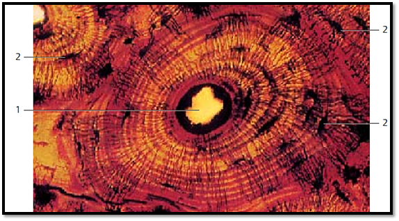
Bone Tissue-Compact Bone
Transverse section of an osteon with its Haversian canal 1 . It is enveloped by lamellae in the ground substance, which may be more or less impregnated with Silver nitrate. The collagen fibers in the more heavily stained lamellae are arrange d in a circular fashion; in the lightly stained areas, their orientation follows the longitudinal axis of the osteons. The black spindle-shaped structures with numerous fine black threads extending from their surfaces are osteocytes 2 and their cellular projections. The osteocytes reside in bone cavities, and their projections reach into the bone canals.
1 Haversian canal
2 Osteocytes
Stain: silver nitrate impregnation; magnification: × 250
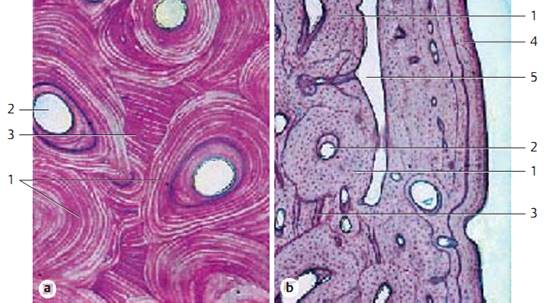
Bone Tissue—Compact Bone
All osteons 1 with their special lamellae and the interstitial lamella systems 3 are encased by the outer and inner general lamellae 4 . Figure b shows the outer general lamella 4 . The oblong narrow space represents a vascularized Volkmann canal ( transverse canal ) 5 . These transverse canals break through the lamella systems. Different lamella systems are separate d by cement lines. These consist of ground substance with a few fibrils, and they stain heavily with hematoxylin.
1 Osteon
2 Haversian canal
3 Interstitial lamella
4 Outer general lamella
5 Volkmann canal
Stain: hematoxylin-eosin; magnifications: a) × 120, b) × 10
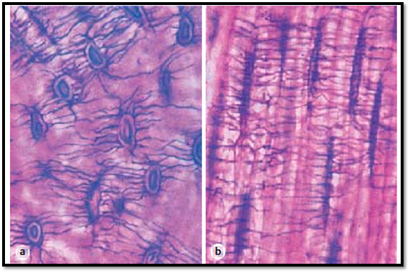
Bone Tissue—Compact Bone
Cross-section (a) and transverse section (b) through an osteon, demonstrating the lacunae (bone cavities, lacunae osseae) and the bone channels (canaliculi ossei). The bone canaliculi cannot be seen in the usual histological preparations. Canaliculi are about 1.0–1.5 μm wide and are devoid of extracellular matrix.
They contain many branched, filopodia-like osteoplast projections. Bone canaliculi of neighboring bone cavities interconnect. The system of canals serves for the exchange of materials between osteocytes and extracellular space. Osteocytes are spindle-shape d cells with long, slender, interconnecting cell projections.
Stain: Schmorl thionine-picric acid; magnification: × 400
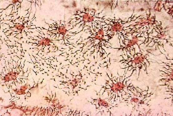
Bone Tissue—Compact Bone
Decalcified bone tissue can be processed like any other tissue. It can be cut using a microtome and stained. The cytoplastic processes of osteoplast occupy bone canals in the extracellular space and connect with each other via nexuses. The exchange of materials probably proceeds via these cytoplasmic processes.Osteocytes and their cytoplasmic processes (projections) are clearly visible in thin sections of compact bone substance from the femur after decalcification and Schmorl staining.
Stain: Schmorl thionine-picric acid; magnification: × 500
References
Kuehnel, W.(2003). Color Atlas of Cytology, Histology, and Microscopic Anatomy. 4th edition . Institute of Anatomy Universitätzu Luebeck Luebeck, Germany . Thieme Stuttgart · New York .
 الاكثر قراءة في علم الخلية
 اخر الاخبار
اخبار العتبة العباسية المقدسة

الآخبار الصحية


مواضيع ذات صلة
 قسم الشؤون الفكرية يصدر كتاباً يوثق تاريخ السدانة في العتبة العباسية المقدسة
 "المهمة".. إصدار قصصي يوثّق القصص الفائزة في مسابقة فتوى الدفاع المقدسة للقصة القصيرة
 (نوافذ).. إصدار أدبي يوثق القصص الفائزة في مسابقة الإمام العسكري (عليه السلام)


