


النبات

مواضيع عامة في علم النبات
الجذور - السيقان - الأوراق
النباتات الوعائية واللاوعائية
البذور (مغطاة البذور - عاريات البذور)
الطحالب
النباتات الطبية
الحيوان
مواضيع عامة في علم الحيوان
علم التشريح
التنوع الإحيائي
البايلوجيا الخلوية
الأحياء المجهرية
البكتيريا
الفطريات
الطفيليات
الفايروسات
علم الأمراض
الاورام
الامراض الوراثية
الامراض المناعية
الامراض المدارية
اضطرابات الدورة الدموية
مواضيع عامة في علم الامراض
الحشرات
التقانة الإحيائية
مواضيع عامة في التقانة الإحيائية
التقنية الحيوية المكروبية
التقنية الحيوية والميكروبات
الفعاليات الحيوية
وراثة الاحياء المجهرية
تصنيف الاحياء المجهرية
الاحياء المجهرية في الطبيعة
أيض الاجهاد
التقنية الحيوية والبيئة
التقنية الحيوية والطب
التقنية الحيوية والزراعة
التقنية الحيوية والصناعة
التقنية الحيوية والطاقة
البحار والطحالب الصغيرة
عزل البروتين
هندسة الجينات
التقنية الحياتية النانوية
مفاهيم التقنية الحيوية النانوية
التراكيب النانوية والمجاهر المستخدمة في رؤيتها
تصنيع وتخليق المواد النانوية
تطبيقات التقنية النانوية والحيوية النانوية
الرقائق والمتحسسات الحيوية
المصفوفات المجهرية وحاسوب الدنا
اللقاحات
البيئة والتلوث
علم الأجنة
اعضاء التكاثر وتشكل الاعراس
الاخصاب
التشطر
العصيبة وتشكل الجسيدات
تشكل اللواحق الجنينية
تكون المعيدة وظهور الطبقات الجنينية
مقدمة لعلم الاجنة
الأحياء الجزيئي
مواضيع عامة في الاحياء الجزيئي
علم وظائف الأعضاء
الغدد
مواضيع عامة في الغدد
الغدد الصم و هرموناتها
الجسم تحت السريري
الغدة النخامية
الغدة الكظرية
الغدة التناسلية
الغدة الدرقية والجار الدرقية
الغدة البنكرياسية
الغدة الصنوبرية
مواضيع عامة في علم وظائف الاعضاء
الخلية الحيوانية
الجهاز العصبي
أعضاء الحس
الجهاز العضلي
السوائل الجسمية
الجهاز الدوري والليمف
الجهاز التنفسي
الجهاز الهضمي
الجهاز البولي
المضادات الميكروبية
مواضيع عامة في المضادات الميكروبية
مضادات البكتيريا
مضادات الفطريات
مضادات الطفيليات
مضادات الفايروسات
علم الخلية
الوراثة
الأحياء العامة
المناعة
التحليلات المرضية
الكيمياء الحيوية
مواضيع متنوعة أخرى
الانزيمات

Eye (Bubble)-Form Intermediate
 المؤلف:
W. L. Fangman and B. J. Brewer
المؤلف:
W. L. Fangman and B. J. Brewer
 المصدر:
Ann. Rev. Cell Biol. 7, 375–402
المصدر:
Ann. Rev. Cell Biol. 7, 375–402
 الجزء والصفحة:
 8-5-2016
 3236
3236


+
-
20
Eye (Bubble)-Form Intermediate
In general, DNA replication of a chromosome starts from a genetically fixed site and proceeds bidirectionally. At the initial stage of replication, the replicated regions form structures like an eye ) or a bubble). This form is important, as it represents the origin region of the chromosome; various methods to detect eye-form intermediates have been developed. Electron microscopy (EM) of the replicating DNA can detect eye-forms as large as several kilobases (1) . Using a combination of digestion by appropriate restriction enzymes and electron microscopy, the origin of replications of plasmids, bacteriophage, and virus DNA has been identified (2). Recently, the two-dimensional gel electrophoresis method was developed to detect various forms of replication intermediates (3). Chromosomal fragments produced by digestion with restriction enzyme are separated by electrophoresis by agarose gel electrophoresis, first by size and then by shape. The distribution of intermediates on the two-dimensional (2D) gel is detected by hybridization with a radiolabeled probe of a cloned DNA fragment from or near the replication origin. Since the correlation between the size and complexity of shape of eye-forms is different from that of Y-fork intermediates, the former can be distinguished from the latter by the shape of arcs on the 2-dimensional gel (Fig. 1). In particular, when the origin of replication is not located at the center of the fragment, the change from eye- to Y-form occurs as replication proceeds on the fragment, which results in a discontinuous distribution of arc from eye- to Y-form. This method has been used successfully to detect the chromosomal origins of eukaryotes, in particular of Saccharomyces cerevisiae (4), Schizosaccharomyces pombe (5), and Drosophila chromosomes (6). Detection of eye-forms from mammalian chromosomes is technically difficult, due to the large amount of background DNA, and this method generally produces ambiguous results as to the site of origin of replication (7.(
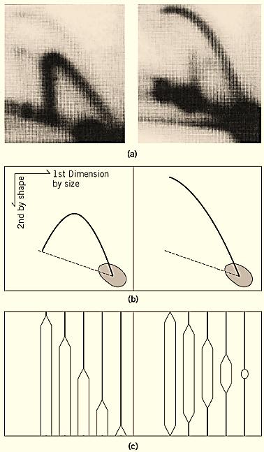
Figure 1. Two-dimensional gel electrophoresis method to detect eye-form (bubble-form) and Y-form replication intermediates. (a) Examples of patterns of Y-form (left) and Eye-form (right) intermediates of the replication of Saccharomyces cerevisiae chromosome are shown. Intermediates were detected by hybridization with the 32P-labeled probe of cloned DNA within the fragments shown in (c). (b) The photos in ( a) are drawn schematically. Dotted lines indicate the location of linear DNA fragments of corresponding size. (c) The population of the intermediates detected in (a) and (b) Left panel: Replication fork moves from the left (or right) side of the fragment and proceeds to produce Y-forks of variable size. Theoretically, a Y-fork with equal branch lengths is structurally most complex and shows least mobility in the second dimension. Right panel: Replication initiates from the center of the fragment and proceeds bidirectionally, producing various sizes of bubble intermediates. The largest bubble is like a circular DNA and shows least mobility in the second dimensions. The locations of the probe used for detection in (a) are indicated by dotted lines. Dark spots in the photos in (a), and drawn schematically in (b), indicate that the majority of DNA detected by the probe is nonreplicated (or completely replicated) linear molecules.
References
1. M. Schnos and R. B. Inman (1970) J. Mol. Biol. 51, 61–73.
2. J. Wolfson, D. Dressler, and M. Magazin (1972) Proc. Natl. Acad. Sci. 69, 499–504.
3. B. J. Brewer and W. L. Fangman (1987) Cell 51, 463–471.
4.W. L. Fangman and B. J. Brewer (1991) Ann. Rev. Cell Biol. 7, 375–402.
5. J. Zhu, D. L. Carlson, D. D. Dubey, K. Sharma, and J. A. Huberman (1994) Chromosoma 103, 414–422 .
6. T. Shinomiya and S. Ina (1991) Nucl. Acids Res. 19, 3935–3941.
7. M. L. DePamphilis (1993) J. Biol. Chem. 268, 1–4.
 الاكثر قراءة في مواضيع عامة في الاحياء الجزيئي
 اخر الاخبار
اخبار العتبة العباسية المقدسة

الآخبار الصحية


مواضيع ذات صلة
 قسم الشؤون الفكرية يصدر كتاباً يوثق تاريخ السدانة في العتبة العباسية المقدسة
 "المهمة".. إصدار قصصي يوثّق القصص الفائزة في مسابقة فتوى الدفاع المقدسة للقصة القصيرة
 (نوافذ).. إصدار أدبي يوثق القصص الفائزة في مسابقة الإمام العسكري (عليه السلام)


