


النبات

مواضيع عامة في علم النبات
الجذور - السيقان - الأوراق
النباتات الوعائية واللاوعائية
البذور (مغطاة البذور - عاريات البذور)
الطحالب
النباتات الطبية
الحيوان
مواضيع عامة في علم الحيوان
علم التشريح
التنوع الإحيائي
البايلوجيا الخلوية
الأحياء المجهرية
البكتيريا
الفطريات
الطفيليات
الفايروسات
علم الأمراض
الاورام
الامراض الوراثية
الامراض المناعية
الامراض المدارية
اضطرابات الدورة الدموية
مواضيع عامة في علم الامراض
الحشرات
التقانة الإحيائية
مواضيع عامة في التقانة الإحيائية
التقنية الحيوية المكروبية
التقنية الحيوية والميكروبات
الفعاليات الحيوية
وراثة الاحياء المجهرية
تصنيف الاحياء المجهرية
الاحياء المجهرية في الطبيعة
أيض الاجهاد
التقنية الحيوية والبيئة
التقنية الحيوية والطب
التقنية الحيوية والزراعة
التقنية الحيوية والصناعة
التقنية الحيوية والطاقة
البحار والطحالب الصغيرة
عزل البروتين
هندسة الجينات
التقنية الحياتية النانوية
مفاهيم التقنية الحيوية النانوية
التراكيب النانوية والمجاهر المستخدمة في رؤيتها
تصنيع وتخليق المواد النانوية
تطبيقات التقنية النانوية والحيوية النانوية
الرقائق والمتحسسات الحيوية
المصفوفات المجهرية وحاسوب الدنا
اللقاحات
البيئة والتلوث
علم الأجنة
اعضاء التكاثر وتشكل الاعراس
الاخصاب
التشطر
العصيبة وتشكل الجسيدات
تشكل اللواحق الجنينية
تكون المعيدة وظهور الطبقات الجنينية
مقدمة لعلم الاجنة
الأحياء الجزيئي
مواضيع عامة في الاحياء الجزيئي
علم وظائف الأعضاء
الغدد
مواضيع عامة في الغدد
الغدد الصم و هرموناتها
الجسم تحت السريري
الغدة النخامية
الغدة الكظرية
الغدة التناسلية
الغدة الدرقية والجار الدرقية
الغدة البنكرياسية
الغدة الصنوبرية
مواضيع عامة في علم وظائف الاعضاء
الخلية الحيوانية
الجهاز العصبي
أعضاء الحس
الجهاز العضلي
السوائل الجسمية
الجهاز الدوري والليمف
الجهاز التنفسي
الجهاز الهضمي
الجهاز البولي
المضادات الميكروبية
مواضيع عامة في المضادات الميكروبية
مضادات البكتيريا
مضادات الفطريات
مضادات الطفيليات
مضادات الفايروسات
علم الخلية
الوراثة
الأحياء العامة
المناعة
التحليلات المرضية
الكيمياء الحيوية
مواضيع متنوعة أخرى
الانزيمات

Plate count method APHA 2001 for Clostridium perfringens in foods
 المؤلف:
SILVA, N.D .; TANIWAKI, M.H. ; JUNQUEIRA, V.C.A.; SILVEIRA, N.F.A. , NASCIMENTO , M.D.D. and GOMES ,R.A.R
المؤلف:
SILVA, N.D .; TANIWAKI, M.H. ; JUNQUEIRA, V.C.A.; SILVEIRA, N.F.A. , NASCIMENTO , M.D.D. and GOMES ,R.A.R
 المصدر:
MICROBIOLOGICAL EXAMINATION METHODS OF FOOD AND WATE A Laboratory Manual
المصدر:
MICROBIOLOGICAL EXAMINATION METHODS OF FOOD AND WATE A Laboratory Manual
 الجزء والصفحة:
 10-3-2016
 7498
7498


+
-
20
Plate count method APHA 2001 for Clostridium perfringens in foods
Method of the American Public Health Association (APHA), as described in the 4th Edition of the Compendium of Methods for the Microbiological Examination of Foods (Labbe, 2001).
1 - Material required for analysis
Preparation of the sample and serial dilutions
• Glycerol-Salt Solution Buffered (to treat samples before storing at 55–60ºC)
• Diluent: 0.1% Peptone Water (PW) or Butterfield’s Phosphate Buffer
• Dilution tubes containing 9 ml of 0.1% Peptone Water (PW) or Butterfield’s Phosphate Buffer
Presumptive counting
• Tryptose Sulfite Cycloserine (TSC) Agar
• Anaerobic jars
• Anaerobic atmosphere generation systems (Anaerogen from Oxoid, Anaerocult A from Merck, GasPak® from BD Biosciences, or equivalent)
• Laboratory incubator set to 35–37°C Confirmation
• Thioglycollate Medium (TGM fluid) (tubes)
• Iron Milk Medium Modified (tubes optional)
• Lactose-Gelatin Medium (tubes)
• Motility-Nitrate Medium (tubes)
• Fermentation Medium for C. perfringens containing 1% salicin (tubes)
• Fermentation Medium for C. perfringens containing
1% reffinose (tubes)
• Nitrate Test Reagents (sulfanilic acid solution, α-naphthol solution)
• 0.04% Bromthymol Blue Indicator
2 - Procedure
A general flowchart for the enumeration of Clostridum perfringens in foods using the plate count method APHA 2001 is shown in Figure 1.
The procedure described below does not present these details, as they are supposed to be known to the analyst.
a) Storage of samples for analysis: The Compendium recommends that samples intended to be used for the enumeration of C. perfringens be analyzed immediately. If impossible, the samples should be refrigerated for the shortest possible time, but should not be frozen. If necessary to store the samples for longer than 48 hours, treat then with Glycerol-Salt Solution Buffered (in an amount required to reach a final concentration of 10% of the sample) and freeze at temperatures between minus 55 and minus 60°C.
b) Preparation of the samples and inoculation .
Since C. perfringens rarely sporulate in food, heat shock for spore count is not recommended.
Select three appropriate dilutions of the sample and inoculate in Tryptose Sulfite Cycloserine (TSC) Agar. Use the pour plating technique and, after complete solidification of the medium, cover the surface with a 5–10 ml thick layer of the same medium. TSC containing egg yolk emulsion may be used (spread plate technique) with an overlay of TSC without egg yolk emulsion.
c) Incubation and colony counting: Incubate the plates (upright position) at 35–37ºC/18–24 h in an anaerobic jar. To establish anaerobic conditions, use anaerobic atmosphere generation systems (Anaerogen from Oxoid, Anaerocult A from Merck, GasPak® from BD Biosciences, or equivalent).
Select plates containing 20 to 200 colonies and count the typical black colonies, which may be sur-rounded by a zone of precipitate on the TSC containing egg yolk.
Caution: TSC allows the growth (and toxin pro-duction) of Clostridium botulinum and its colonies cannot be distinguished from C. perfringens colonies (Serrano & Junqueira, 1991). The plates and all the cultures isolated during confirmation should be handled with care.
d) Confirmation: Select five typical colonies for confirmation and if there are fewer than five, select all. Transfer the suspect colonies to Thioglycollate Medium (TGM) tubes (before inoculation exhaust oxygen from the tubes). Incubate the tubes at 46°C/4 h (water bath) or at 35–37°C/18–20 h.
Check for purity (Gram stain) and purify if contaminated (streak on TSC, incubate at 35–37ºC/18–24 h in anaerobic condition, transfer a well isolated colony to TGM and incubate TGM at 46°C/4 h or at 35–37°C/18–20 h). Use the TGM cultures for confirmatory tests.
d.1) Lactose fermentation test and gelatin liquefaction test: From each culture, inoculate a tube of Lactose-Gelatin Medium by stabbing (exhaust oxygen from tubes before inoculation). Incubate the tubes at 35–37°C/24–44 h. A color change from red to yellow indicates a positive reaction for lactose fermentation and the lack of color change indicates a negative reaction. Refrigerate the tubes at 5°C/2 h and examine for gelatin solidification. If the medium remains liquid, the reaction is positive (gelatin liquefied). If the medium solidifies (negative result), incubate an additional 24 h at 35°C and test again. C. perfringens strains ferment lactose and liquefy gelatin.
d.2) Motility test and nitrate reduction test: From each culture, inoculate a tube of Motility-Nitrate Medium by stabbing (exhaust oxygen from tubes before inoculation). Incubate the tubes at 35–37°C/24 h and check for motility. C. perfringens is non-motile and the growth will occur only along the line of inoculum. To test for nitrate reduction to
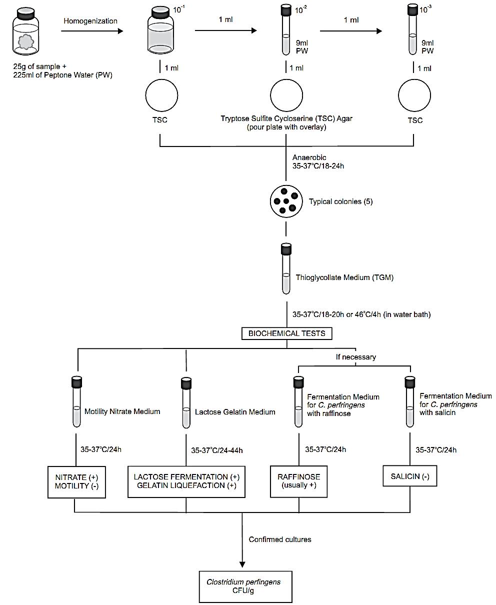
Figure 1 Scheme of analysis for the enumeration of Clostridium perfringens in foods using the plate count method APHA 2001 (Labbe, 2001).
nitrite, add 0.5 ml each of nitrate test reagents (sulfanilic acid solution and α-naphthol solution) to each culture. An orange color indicates a positive reaction (nitrite present, nitrate reduced to nitrite). If no color develops (nitrite absent), test for residual nitrate by adding a small amount of zinc dust. An orange color indicates a negative reaction (nitrate is present, has not been reduced) and the absence of color indicates a positive reaction (nor nitrate or nitrite present, nitrate has been completely reduced to N2). C. perfringens strains are positive for nitrate reduction.
d.3) Raffinose and salicin fermentation tests: The raffinose and salicin fermentation tests are required only for cultures that do not liquefy gelatin within 44 h, or are atypical in other respects. Use a 24 h TGM culture for the tests. Inoculate 0.1 ml of TGM culture into a tube of Fermentation Medium for C. perfringens containing 1% salicin and 0.1 ml into a tube of the same medium containing 1% raffinose (exhaust oxygen from tubes before inoculation). Incubate the tubes at 35–37°C/24 h and check for acid production. To test for acid, transfer 1 ml of the culture to a test tube and add two drops of a 0.04% Bromthymol Blue Indicator. A yellow color indicates that acid has been produced. Reincubate negative cultures for an additional 48 h and retest for acid production. C. perfringens strains usually ferment raffinose and do not ferment salicin.
e) Calculation of results: Consider as C. perfringens the cultures of non-motile Gram positive rods showing lactose fermentation positive, gelatin liquefaction positive, and nitrate reduction positive. If one of these reactions is atypical, consider the results of raffinose fermentation (usually positive), salicin fermentation (usually negative) and “stormy fermentation” (positive) to consider the cultures as C. perfringens.
Calculate number of cells/g of sample based on percentage of colonies tested that are confirmed as C. perfringens.
Example 1: The presumptive count obtained with 10−4 dilution of sample was 65 (pour plate, 1 ml inoculated). Four of five colonies tested were con-firmed (80%). The number of C. perfringens cells/g of food is 65 × 0.8 × 104 = 5.2 × 10⁵ CFU/g.
Example 2: The presumptive count obtained with 10−3 dilution of sample was 30 (spread plate, 0.1 ml inoculated). Two of ten colonies tested were con-firmed (20%). The number of C. perfringens cells/g of food is 30 × 0.2 × 103 × 10 = 6.0 × 10⁴ CFU/g (dilution factor is tenfold higher than sample dilution because only 0.1 ml was tested).
References
Silva, N.D .; Taniwaki, M.H. ; Junqueira, V.C.A.; Silveira, N.F.A. , Nasdcimento , M.D.D. and Gomes ,R.A.R .(2013) . Microbiological examination methods of food and water a laboratory Manual. Institute of Food Technology – ITAL, Campinas, SP, Brazil .
Labbe, R.G. (2001) Clostridium perfringens. In: Downes, F.P. & Ito, K. (eds.). Compendium of Methods for the Microbiological Examination of Foods. 4th edition. Washington, American Public Health Association. Chapter 34, pp. 325–330.
FDA/CFSAN (ed.) (2009) Foodborne Pathogenic Microorganisms and Natural Toxins Handbook “Bad Bug Book”. [Online] College Park, Food and Drug Administration, Center for Food Safety & Applied Nutrition. Available from: http://www.fda.gov/food/foodsafety/foodborneillness/foodborneillnessfoodbornepa-thogensnaturaltoxins/badbugbook/default.htm [accessed 10th October 2011].
Murrell, T.G.C. (1983). Pigbel in Papua New Guinea: An Ancient Disease Rediscovered. International Journal of Epidemiology, 12(2), 211–214.
 الاكثر قراءة في البكتيريا
 اخر الاخبار
اخبار العتبة العباسية المقدسة

الآخبار الصحية


مواضيع ذات صلة
 قسم الشؤون الفكرية يصدر كتاباً يوثق تاريخ السدانة في العتبة العباسية المقدسة
 "المهمة".. إصدار قصصي يوثّق القصص الفائزة في مسابقة فتوى الدفاع المقدسة للقصة القصيرة
 (نوافذ).. إصدار أدبي يوثق القصص الفائزة في مسابقة الإمام العسكري (عليه السلام)


