Catalase
Catalase, also called hydroperoxidase, is a protective enzyme that has been studied for over a century; the concept of a specific protein catalyzing (hence the name catalase) the degradation of hydrogen peroxide to oxygen and water first appeared in 1900 (1). Removal of H2O2, as shown in the following reaction, avoids unwanted side reactions and prevents the formation of the even more reactive hydroxyl radical:

Although the removal of these reactive oxygen species is not essential for growth, catalases do enhance long-term survival in an aerobic environment (2).
A number of developments have spawned considerable interest in the enzyme and spurred extensive studies of both the enzyme and its genes. The first was the simplicity of the “drop test” assay, which involves the visual monitoring of oxygen evolution after the application of a drop of 30% H2O2 to the edge of a bacterial colony. Such ease of phenotypic scoring has resulted in extensive use of the enzyme as a diagnostic tool for microbiological strain identification. Catalases have been found to be synthesized as part of a variety of stress response systems, resulting in their frequent use as indicators of stress response activation. More recently, the catalase–peroxidase family has gained notoriety from its role as the in vivo activator of isoniazid into an effective antibiotic in Mycobacterium tuberculosis (3). Loss of the enzyme through mutation results in isoniazid resistance, one of the reasons for the increasing spread of tuberculosis.
1. Assay
Two quantitative assay methods are commonly used for catalase activity. One involves the measurement of oxygen evolution using an oxygraph equipped with a Clark electrode (4); the second is a spectrophotometric assay of H2O2 by its absorbance at 240 nm (5). The two assay procedures produce comparable values in reasonably clear protein solutions, but the oxygraph protocol has the advantage that it can be used for catalase determinations in whole-cell suspensions and in quite turbid extracts. Catalase activity can be visualized on polyacrylamide gels following electrophoresis under nondenaturing conditions (6), producing a clear band in a brown background. This same visualization procedure can be easily modified to visualize peroxidase activities as brown bands on a clear background.
2. Classification
Because of the protection afforded against active oxygen species by catalases, most aerobic organisms produce at least one catalase from among the three main classes of the enzyme. The largest class includes the “typical,” heme-containing, monofunctional catalases with either small ~60 (kDa) or large (>80 kDa) subunits. The next largest class includes the bifunctional catalase–peroxidases, which are also heme-containing but with sequence similarity to plant and fungal peroxidases. The third and, to date, smallest class includes the non-heme- or Mn-containing catalases. Small-subunit, monofunctional catalases have been found in prokaryotes and eukaryotes but not in an archael species, and the large-subunit catalases are restricted to fungi and bacteria. The catalase-peroxidases are found in prokaryotes and archaebacteria, and the non-heme-containing enzymes have been found only in bacteria (7).
3. Reaction
Catalases and peroxidases both degrade H2O2 but, whereas catalase employs H2O2 as both electron donor and acceptor (reaction 1), a peroxidase uses organic substrates as the electron donor to reduce H2O2:

The catalase-peroxidases catalyze both reactions 1 and 2 at significant rates.
The well-characterized catalytic reaction pathway is a two-step process. First, H2O2 is converted to water and an oxyferryl species, compound I, with the iron in the +5 oxidation state but with part of the charge delocalized in a heme cation radical:

Then compound I reacts with a second molecule of H2O2 to produce water and molecular oxygen:

In the absence of a suitable substrate for the reduction of compound I, other intermediates can be formed such as the catalytically inactive compound II.
4. Structure and Properties
Detailed structural information is now available for all three types of catalases. Small- and large-subunit catalases share a common highly conserved core sequence of about 350 residues organized in a b-barrel structure. This is illustrated in Figure 1 (8, 9), which compares the small subunit of Proteus mirabilis catalase (Fig. 1a) and the large subunit of Escherichia coli HPII (Fig. 1b). An N-terminal extension of 10–90 residues (colored red in Fig. 1B) and a C-terminal extension of 150–170 residues (colored white in Fig. 1b), the latter organized in a flavodoxin-like structure, are added to the core structure in the large-subunit enzyme. The tetrameric organization of small-subunit enzymes is sufficiently stable to resist denaturation by organic reagents and to retain activity over a broad pH range from 4 to 11. In large-subunit enzymes, the extended sequences allow the amino terminus of one subunit to be overlapped or trapped by the carboxyl terminus of an adjacent subunit (10), and this interweaving further enhances the resistance of the tetramer to denaturation by heat, SDS, and urea.
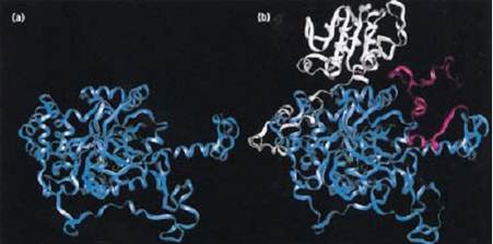
Figure 1. Comparison of the structures of the small subunit of Proteus mirabilis catalase (a) (8) and the large subunit of E. coli HPII catalase (b) (9). The conserved b-barrel core structure is colored blue in both subunits. The 78 additional residues at the N terminus of HPII in (b) are colored red, and the 195 additional residues at the C terminus of HPII in (b) are colored white.
Besides size, the small- and large-subunit catalases have two other characteristic differences. The first is that small-subunit enzymes contain heme b, which in large-subunit enzymes is converted to a cis-hydroxy g-spirolactone heme d and flipped 180°. The second is that small-subunit, but not large-subunit, catalases contain NADPH (11), which is postulated to have a role in the reduction of inactive compound II. A number of unusual modifications have been identified in catalases, including
1. A methionine sulfone in the active site of the P. mirabilis enzyme (12)
2. A blocked cysteine residue in HPII from E. coli (13(
3. A histidine–tyrosine bond in HPII
The narrow channel leading to the deeply buried active site of catalases limits access to small substrates and inhibitors, and results in a 10-fold higher apparent Km for H2O2 compared to the non-heme-containing catalases and catalase–peroxidases. Within the catalase active site, a histidine residue immediately above the heme, and an asparagine residue situated to one side, orient the H2O2 over the heme iron (12) to facilitate a very fast reaction in both small- and large-subunit enzymes [kcat @ 3.5 × 10⁵ s–1 (15) and 1.2 × 10⁵ s–1 , respectively] as compared to the catalase–peroxidases and non-heme-containing catalases [kcat @ 1.6 × 10⁴ s–1 (16) and 3.9 × 10⁵ s–1 (17), respectively.
5. Cloning and Expression
The number of catalase enzymes in a given organism varies: one in animals, one or two in fungi, one to three in plants, and zero to three in bacteria. The availability of catalase-deficient hosts, the ease of assay, and the development of protocols using oligonucleotide and PCR probes have facilitated the cloning and characterization of over 90 catalase genes. Most of the isolated genes are from plants and bacteria, with a small number from fungi and animals. A phylogenetic comparison of the core protein sequences has revealed separate branches for the small-subunit enzymes from bacterial, plant, animal, and fungal sources, and one additional branch containing the large-subunit enzymes from both fungal and bacterial origins.
The most detailed studies of catalase gene expression have been carried out in plants and bacteria. In maize, catalase expression is tied to developmental changes during kernel development and seed germination and to environmental factors such as light and growth regulators (18). A multitude of species-specific mechanisms control catalase and catalase–peroxidase expression in bacteria, but two common threads are evident among these expression patterns involving induction either by an active oxygen species such as H2O2 or in stationary phase. In most cases, the control mechanisms can be rationalized as either protective responses to oxidative stress or as a means of enhancing long-term survival during metabolic limitation (7, 19).
References
1. O. Loew (1900) U.S. Dept. Agriculture Rpt. 65, Govt. Printing Office, Washington, DC.
2. M. R. Mulvey, J. Switala, A. Borys, and P. C. Loewen (1990) J. Bacteriol. 172, 6713–6720.
3. J. S. Blanchard (1996) Annu. Rev. Biochem. 65, 215–239.
4. M. Rorth and P. K. Jensen. (1967) Biochim. Biophys. Acta 139, 171–173.
5. A. G. Hildebrandt and I. Roots (1975) Arch. Biochem. Biophys. 171, 385–397.
6. E. M. Gregory and I. Fridovich. (1974) Anal. Biochem. 58, 57–62.
7. P. C. Loewen (1997) In J. G. Scandalios, ed., Oxidative Stress and the Molecular Biology of Antioxidant Defenses, Cold Spring Harbor Laboratory Press, Cold Spring Harbor, NY, pp. 273–308 .
8. P. Gouet, H. M. Jouve, and O. Dideberg (1995) J. Mol. Biol. 249, 933–954.
9. J. Bravo, N. Verdaguer, J. Tormo, C. Betzel, J. Switala, P. C. Loewen, and I. Fita (1995( Structure 3, 491–502.
10. W. R. Melik-Adamyan, V. V. Barynin, A. A. Vagin, V. V. Borisov, B. K. Vainshtein, I. Fita, M. R. N. Murthy, and M. G. Rossmann (1986) J. Mol. Biol. 188, 63–72.
11. H. N. Kirkman and G. F. Gaetani (1984) Proc. Natl. Acad. Sci. USA 81, 4343–4347.
12. A. Buzy, V. Bracchi, R. Sterjiades, J. Chroboczek, P. Thibault, J. Gagnon, H.-M. Jouve, and G. Hudry-Clergeon (1995) J. Protein Chem. 14, 59–72.
13. S. Sevinc, W. Ens, and P. C. Loewen (1995) Eur. J. Biochem. 230, 127–132.
14. I. Fita and M. G. Rossmann (1985) J. Mol. Biol. 185, 21–37.
15. A. Deisseroth and A. L. Dounce (1970) Physiol. Rev. 50, 319–375.
16.A. Claiborne and I. Fridovich (1979) J. Biol. Chem. 254, 4245–4252.
17. Y. Kono and I. Fridovich (1983) J. Biol. Chem. 258, 6015–6019.
18. J. G. Scandalios (1992) in J. G. Scandalios, ed., Molecular Biology of Free Radical Scavenging Systems, Cold Spring Harbor Laboratory Press, Cold Spring Harbor, NY, pp. 117–152.
19. H. Schellhorn (1994) FEMS Microbiol. Lett. 131, 113–119.


