


النبات

مواضيع عامة في علم النبات
الجذور - السيقان - الأوراق
النباتات الوعائية واللاوعائية
البذور (مغطاة البذور - عاريات البذور)
الطحالب
النباتات الطبية
الحيوان
مواضيع عامة في علم الحيوان
علم التشريح
التنوع الإحيائي
البايلوجيا الخلوية
الأحياء المجهرية
البكتيريا
الفطريات
الطفيليات
الفايروسات
علم الأمراض
الاورام
الامراض الوراثية
الامراض المناعية
الامراض المدارية
اضطرابات الدورة الدموية
مواضيع عامة في علم الامراض
الحشرات
التقانة الإحيائية
مواضيع عامة في التقانة الإحيائية
التقنية الحيوية المكروبية
التقنية الحيوية والميكروبات
الفعاليات الحيوية
وراثة الاحياء المجهرية
تصنيف الاحياء المجهرية
الاحياء المجهرية في الطبيعة
أيض الاجهاد
التقنية الحيوية والبيئة
التقنية الحيوية والطب
التقنية الحيوية والزراعة
التقنية الحيوية والصناعة
التقنية الحيوية والطاقة
البحار والطحالب الصغيرة
عزل البروتين
هندسة الجينات
التقنية الحياتية النانوية
مفاهيم التقنية الحيوية النانوية
التراكيب النانوية والمجاهر المستخدمة في رؤيتها
تصنيع وتخليق المواد النانوية
تطبيقات التقنية النانوية والحيوية النانوية
الرقائق والمتحسسات الحيوية
المصفوفات المجهرية وحاسوب الدنا
اللقاحات
البيئة والتلوث
علم الأجنة
اعضاء التكاثر وتشكل الاعراس
الاخصاب
التشطر
العصيبة وتشكل الجسيدات
تشكل اللواحق الجنينية
تكون المعيدة وظهور الطبقات الجنينية
مقدمة لعلم الاجنة
الأحياء الجزيئي
مواضيع عامة في الاحياء الجزيئي
علم وظائف الأعضاء
الغدد
مواضيع عامة في الغدد
الغدد الصم و هرموناتها
الجسم تحت السريري
الغدة النخامية
الغدة الكظرية
الغدة التناسلية
الغدة الدرقية والجار الدرقية
الغدة البنكرياسية
الغدة الصنوبرية
مواضيع عامة في علم وظائف الاعضاء
الخلية الحيوانية
الجهاز العصبي
أعضاء الحس
الجهاز العضلي
السوائل الجسمية
الجهاز الدوري والليمف
الجهاز التنفسي
الجهاز الهضمي
الجهاز البولي
المضادات الميكروبية
مواضيع عامة في المضادات الميكروبية
مضادات البكتيريا
مضادات الفطريات
مضادات الطفيليات
مضادات الفايروسات
علم الخلية
الوراثة
الأحياء العامة
المناعة
التحليلات المرضية
الكيمياء الحيوية
مواضيع متنوعة أخرى
الانزيمات

Peripheral nerves
 المؤلف:
Stuart H. Ralston , Ian D Penman, Mark W J Strachan , Richard Hobson
المؤلف:
Stuart H. Ralston , Ian D Penman, Mark W J Strachan , Richard Hobson
 المصدر:
Davidsons Principles and Practice of Medicine
المصدر:
Davidsons Principles and Practice of Medicine
 الجزء والصفحة:
24th Edition , p156-157
الجزء والصفحة:
24th Edition , p156-157
 2025-02-15
 1055
1055


+
-
20
Peripheral nerves contain several types of neuron. These can be classified into two groups, depending on whether or not they are surrounded by a myelin sheath. Myelinated neurons have a fast conduction velocity and are responsible for transmission of various sensory signals, such as proprioception, light touch, heat and cold, and the detection of localised pains, such as pin-prick. Unmyelinated fibres have a much slower con duction velocity and are responsible for transmitting diffuse and poorly localised pain, as well as other sensations (Box 1).
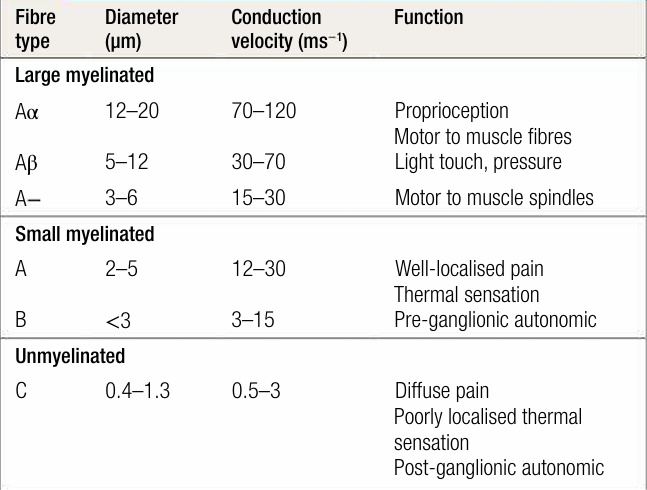
Box1. Types of nerve fibre
Sensory neurons (also known as primary afferent neurons) connect the spinal cord to the periphery and supply a defined territory or a dermatome, which can be used to identify the position of a nerve lesion ( Fig. 1). In healthy individuals, dermatomes have distinct borders, but in pathological pain syndromes these may become blurred as the result of neuronal plasticity, which means that pain may be felt in an area adjacent to that supplied by a specific nerve root. Autonomic neurons also contain pain fibres and are responsible for transmitting visceral sensations, such as colic. In general, visceral pain is diffuse and less well localised than pain transmitted by sensory neurons.

fig1. The areas supplied by specific levels of the spinal cord.
Anatomical features of the afferent pain pathway are illustrated in Figure 2. Pain signals are transmitted from the periphery to the spinal cord by sensory neurons. These have the following components:
- A cell body, containing the nucleus, which is situated in the dorsal root ganglion close to the spinal cord. The cell body is essential for survival of the neuron, production of neurotransmitters and neuronal function.
- The nerve fibre (axon) and peripheral nerve endings, which are located in the periphery and contain a range of receptors in the neuronal membrane.
- Specialised receptors in the periphery, consisting of bare nerve endings known as nociceptors or pain receptors, which are activated by various mediators. They are situated mainly in the epidermis.
- The central termination, which travels to the dorsal horn of the spinal cord to form the first central synapse with neurons that transmit pain sensation to the brain.
When a noxious stimulus is encountered, activation of nociceptors leads to generation of an action potential, which travels upwards to the dorsal root ganglion and also stimulates the release of neurotransmitters that have secondary effects on surrounding neurons.
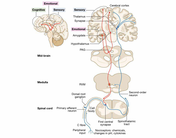
fig2. Ascending and descending pain pathways.
 الاكثر قراءة في الجهاز العصبي
 اخر الاخبار
اخبار العتبة العباسية المقدسة

الآخبار الصحية


مواضيع ذات صلة
 قسم الشؤون الفكرية يصدر كتاباً يوثق تاريخ السدانة في العتبة العباسية المقدسة
 "المهمة".. إصدار قصصي يوثّق القصص الفائزة في مسابقة فتوى الدفاع المقدسة للقصة القصيرة
 (نوافذ).. إصدار أدبي يوثق القصص الفائزة في مسابقة الإمام العسكري (عليه السلام)


