


النبات

مواضيع عامة في علم النبات
الجذور - السيقان - الأوراق
النباتات الوعائية واللاوعائية
البذور (مغطاة البذور - عاريات البذور)
الطحالب
النباتات الطبية
الحيوان
مواضيع عامة في علم الحيوان
علم التشريح
التنوع الإحيائي
البايلوجيا الخلوية
الأحياء المجهرية
البكتيريا
الفطريات
الطفيليات
الفايروسات
علم الأمراض
الاورام
الامراض الوراثية
الامراض المناعية
الامراض المدارية
اضطرابات الدورة الدموية
مواضيع عامة في علم الامراض
الحشرات
التقانة الإحيائية
مواضيع عامة في التقانة الإحيائية
التقنية الحيوية المكروبية
التقنية الحيوية والميكروبات
الفعاليات الحيوية
وراثة الاحياء المجهرية
تصنيف الاحياء المجهرية
الاحياء المجهرية في الطبيعة
أيض الاجهاد
التقنية الحيوية والبيئة
التقنية الحيوية والطب
التقنية الحيوية والزراعة
التقنية الحيوية والصناعة
التقنية الحيوية والطاقة
البحار والطحالب الصغيرة
عزل البروتين
هندسة الجينات
التقنية الحياتية النانوية
مفاهيم التقنية الحيوية النانوية
التراكيب النانوية والمجاهر المستخدمة في رؤيتها
تصنيع وتخليق المواد النانوية
تطبيقات التقنية النانوية والحيوية النانوية
الرقائق والمتحسسات الحيوية
المصفوفات المجهرية وحاسوب الدنا
اللقاحات
البيئة والتلوث
علم الأجنة
اعضاء التكاثر وتشكل الاعراس
الاخصاب
التشطر
العصيبة وتشكل الجسيدات
تشكل اللواحق الجنينية
تكون المعيدة وظهور الطبقات الجنينية
مقدمة لعلم الاجنة
الأحياء الجزيئي
مواضيع عامة في الاحياء الجزيئي
علم وظائف الأعضاء
الغدد
مواضيع عامة في الغدد
الغدد الصم و هرموناتها
الجسم تحت السريري
الغدة النخامية
الغدة الكظرية
الغدة التناسلية
الغدة الدرقية والجار الدرقية
الغدة البنكرياسية
الغدة الصنوبرية
مواضيع عامة في علم وظائف الاعضاء
الخلية الحيوانية
الجهاز العصبي
أعضاء الحس
الجهاز العضلي
السوائل الجسمية
الجهاز الدوري والليمف
الجهاز التنفسي
الجهاز الهضمي
الجهاز البولي
المضادات الميكروبية
مواضيع عامة في المضادات الميكروبية
مضادات البكتيريا
مضادات الفطريات
مضادات الطفيليات
مضادات الفايروسات
علم الخلية
الوراثة
الأحياء العامة
المناعة
التحليلات المرضية
الكيمياء الحيوية
مواضيع متنوعة أخرى
الانزيمات

Chromatin Is Divided into Euchromatin and Heterochromatin
 المؤلف:
JOCELYN E. KREBS, ELLIOTT S. GOLDSTEIN and STEPHEN T. KILPATRICK
المؤلف:
JOCELYN E. KREBS, ELLIOTT S. GOLDSTEIN and STEPHEN T. KILPATRICK
 المصدر:
LEWIN’S GENES XII
المصدر:
LEWIN’S GENES XII
 الجزء والصفحة:
 22-3-2021
 5090
5090


+
-
20
Chromatin Is Divided into Euchromatin and Heterochromatin
KEY CONCEPTS
- We can see individual chromosomes only during mitosis.
- During interphase, the general mass of chromatin is in the form of euchromatin, which is slightly less tightly packed than mitotic chromosomes.
- Regions of heterochromatin remain densely packed throughout interphase.
Each chromosome contains a single, very long duplex of DNA, folded into a fiber that runs continuously throughout the chromosome. Thus, in accounting for interphase chromatin and mitotic chromosome structure, we have to explain the packaging of a single, exceedingly long molecule of DNA into a form in which it can be transcribed and replicated, and can become cyclically more and less compressed.
Individual eukaryotic chromosomes become visible as single compact units during mitosis. FIGURE 1 is an electron micrograph of a replicated chromosome isolated and photographed at metaphase. The sister chromatids are evident at this stage, and will give rise to the daughter chromosomes upon their separation starting at anaphase. Each chromatid consists of a large thick fiber with a nubbly appearance. The DNA is 5 to 10 times more condensed in mitotic chromosomes than in interphase chromatin.
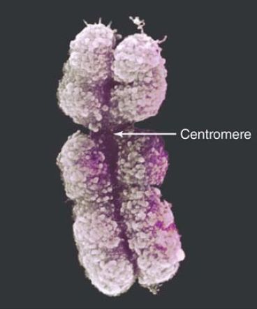
FIGURE 1. The sister chromatids of a mitotic pair each consist of a fiber (~30 nm in diameter) compactly folded into the chromosome.
© Biophoto Associates/Science Source.
During most of the life cycle of the eukaryotic cell, however, its genetic material occupies an area of the nucleus in which individual chromosomes cannot be distinguished by conventional microscopy. The global structure of the interphase chromatin does not appear to change visibly between divisions or even during the period of replication, when the amount of chromatin doubles. Chromatin is fibrillar, although the overall spatial configuration of the fiber has long been difficult to discern. However, recent advances in highresolution microscopy, fluorescence in situ hybridization (FISH) staining, and live imaging have finally begun to reveal additional aspects of chromatin structure and nuclear architecture not evident in the last century.
As the nuclear section of FIGURE 2 illustrates, we can divide chromatin into two types of material:
- In most regions, the chromatin is less densely packed than in the mitotic chromosome. This material, called euchromatin, is relatively dispersed and occupies most of the nucleoplasm.
- Some regions of chromatin are very densely packed, displaying a condition comparable to that of the chromosome at mitosis. This material, called heterochromatin, is typically found at centromeres, but occurs at other locations as well, including telomeres and highly repetitive sequences. It passes through the cell cycle with relatively little change in its degree of condensation. It forms a series of discrete clumps, visible in Figure 2, with a tendency to be found at the nuclear periphery and at the nucleolus. In some cases, the various heterochromatic regions, especially those associated with centromeres, aggregate into a densely staining chromocenter.
The common form of heterochromatin that always remains heterochromatic is called constitutive heterochromatin. In contrast, there is another category of heterochromatin, called facultative heterochromatin, in which regions of euchromatin are converted to a heterochromatic state.
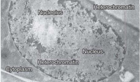
FIGURE 2 A thin section through a nucleus stained with Feulgen shows heterochromatin as compact regions clustered near the nucleolus and nuclear membrane.
Photo courtesy of Edmund Puvion, Centre National de la Recherche Scientifique.
The same fibers run continuously between euchromatin and heterochromatin, as these states simply represent different degrees of condensation of the genetic material. In the same way, euchromatic regions exist in different states of condensation during interphase and mitosis. Thus, the genetic material is organized in a manner that permits alternative states to be maintained side by side in chromatin, and allows cyclical changes to occur in the packaging of euchromatin between interphase and division. We discuss the molecular basis for these states in the chapters titled Chromatin and Epigenetics I and II.
The structural condition of the genetic material is correlated with its activity. The common features of constitutive heterochromatin are as follows:
-It is permanently or nearly always condensed.
-It replicates late in S phase and has a reduced frequency of genetic recombination relative to euchromatic gene-rich areas of the genome.
-It often consists of multiple repeats of a few sequences of DNA that are not transcribed or are transcribed at very low levels. (Genes that reside in heterochromatic regions are generally less transcriptionally active than their euchromatic counterparts, but there are exceptions to this general rule.)
-The density of genes in this region is very much reduced compared with euchromatin, and genes that are translocated into or near it are often inactivated. The one dramatic exception to this is the ribosomal DNA in the nucleolus, which has the general compacted appearance and behavior of heterochromatin (such as late replication), yet is engaged in very active transcription.
There are numerous molecular markers for changes in the properties of the DNA and protein components . They include reduced acetylation of histone proteins, increased methylation at particular sites on histones, and methylation of cytosine bases in DNA. These molecular changes result in the condensation of the chromatin and the recruitment of heterochromatin-specific proteins, which are responsible for maintaining or spreading its inactivity. Although active genes are contained within euchromatin, only a minority of the sequences in euchromatin are transcribed at any time. Thus, location in euchromatin is necessary for most gene expression, but is not sufficient for it.
In addition to the general distributions observed for heterochromatin and euchromatin, studies have addressed whether there is an overall chromosome organization within the nucleus. The answer in many cases is yes; chromosomes appear to occupy distinct three-dimensional spaces known as chromosome territories, as diagrammed in FIGURE 3, showing a probabilistic model of the spatial arrangement of human chromosome territories. The chromosomes occupying these territories are not entangled with one another, but do share areas of interaction and some common functional organization. For example, heterochromatic and other silent regions are found primarily at the nuclear periphery, whereas gene-dense regions are internally located. Active genes are often found at the borders of territories, sometimes clustered together in interchromosomal spaces that are enriched in transcriptional machinery, known as transcription factories.
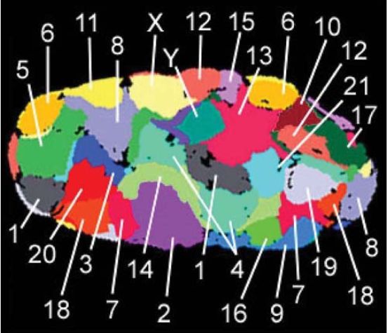
FIGURE 3 Chromosomes occupy chromosome territories in the nucleus and are not entangled with one another. This is a falsecolored representation of chromosome territories obtained by individually staining chromosomes 1–22, X and Y in a human fibroblast nucleus. Heterochromatic regions, silenced genes, and gene-sparse regions of chromosomes are typically localized to the nuclear periphery. Active genes are often found at the borders of chromosome territories, and active genes from several chromosomes can cluster in interchromosomal territories that are enriched in transcription machinery.
Data from Bolzer, A., et al. 2005. PLoS Biol 3(5): e157.
How chromosome territories are established, and how they vary by cell cycle and cell type, are not yet understood, but advances in super-resolution microscopy, genomics, and mathematical modeling are beginning to reveal the presence of subchromosomal compartments and domains that occur in the historically refractory structural scale between a 30-nm chromatin fiber and whole chromosomes. For instance, researchers can define large chromosomal domains by the time at which they replicate in S phase. Comparing replication-timing profiles of several mammalian cell types reveals that the changes occur in defined units of 400–800 kb called replication domains (RDs). As summarized in FIGURE 4, these RDs correspond to structural domains called topologically associated domains (TADs), as revealed by chromatin interaction maps described in the Chromatin chapter. Evidence for this relationship comes from the concomitant switching between RDs and TAD compartments as cells differentiate. In this regard, RDs and TADs might represent chromosomal subdomains or nuclear compartments that act as epigenetic modules preserved across cell types.
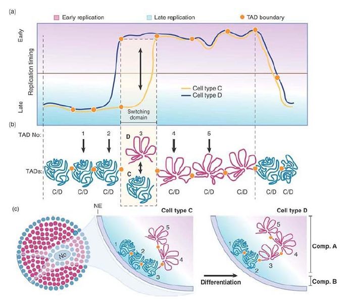
FIGURE 4 Chromatin is regulated at the level of defined units during differentiation. (a) Changes in temporal order of replication timing identify units of chromosome structure. Comparing replication timing profiles of two hypothetical cell types (C and D) identifies a replication domain that change replication timing during differentiation (switching domain). (b) Replication domains correspond to TADs. TADs can be early replicating and open (red) or late replicating and closed (green) depending on the cell type. Exemplary TADs are numbered 1 to 5. TADs 1 and 2 are late replicating, and TADs 4 and 5 are early replicating in both cell types. TAD 3 is late replicating in cell type C and early replicating in cell type D. (c) In general, early replicating TADs (red circles) are more open and located in the nuclear interior, and late replicating TADs (green circles) are more compact and located toward the nuclear periphery. During differentiation, TADs that switch replication timing move toward or away from nuclear lamina and undergo a change in compaction depending on the direction of the replicating timing switch.
 الاكثر قراءة في مواضيع عامة في الاحياء الجزيئي
 اخر الاخبار
اخبار العتبة العباسية المقدسة

الآخبار الصحية


مواضيع ذات صلة
 قسم الشؤون الفكرية يصدر كتاباً يوثق تاريخ السدانة في العتبة العباسية المقدسة
 "المهمة".. إصدار قصصي يوثّق القصص الفائزة في مسابقة فتوى الدفاع المقدسة للقصة القصيرة
 (نوافذ).. إصدار أدبي يوثق القصص الفائزة في مسابقة الإمام العسكري (عليه السلام)


