
النبات

مواضيع عامة في علم النبات

الجذور - السيقان - الأوراق

النباتات الوعائية واللاوعائية

البذور (مغطاة البذور - عاريات البذور)

الطحالب

النباتات الطبية


الحيوان

مواضيع عامة في علم الحيوان

علم التشريح

التنوع الإحيائي

البايلوجيا الخلوية


الأحياء المجهرية

البكتيريا

الفطريات

الطفيليات

الفايروسات


علم الأمراض

الاورام

الامراض الوراثية

الامراض المناعية

الامراض المدارية

اضطرابات الدورة الدموية

مواضيع عامة في علم الامراض

الحشرات


التقانة الإحيائية

مواضيع عامة في التقانة الإحيائية


التقنية الحيوية المكروبية

التقنية الحيوية والميكروبات

الفعاليات الحيوية

وراثة الاحياء المجهرية

تصنيف الاحياء المجهرية

الاحياء المجهرية في الطبيعة

أيض الاجهاد

التقنية الحيوية والبيئة

التقنية الحيوية والطب

التقنية الحيوية والزراعة

التقنية الحيوية والصناعة

التقنية الحيوية والطاقة

البحار والطحالب الصغيرة

عزل البروتين

هندسة الجينات


التقنية الحياتية النانوية

مفاهيم التقنية الحيوية النانوية

التراكيب النانوية والمجاهر المستخدمة في رؤيتها

تصنيع وتخليق المواد النانوية

تطبيقات التقنية النانوية والحيوية النانوية

الرقائق والمتحسسات الحيوية

المصفوفات المجهرية وحاسوب الدنا

اللقاحات

البيئة والتلوث


علم الأجنة

اعضاء التكاثر وتشكل الاعراس

الاخصاب

التشطر

العصيبة وتشكل الجسيدات

تشكل اللواحق الجنينية

تكون المعيدة وظهور الطبقات الجنينية

مقدمة لعلم الاجنة


الأحياء الجزيئي

مواضيع عامة في الاحياء الجزيئي


علم وظائف الأعضاء


الغدد

مواضيع عامة في الغدد

الغدد الصم و هرموناتها

الجسم تحت السريري

الغدة النخامية

الغدة الكظرية

الغدة التناسلية

الغدة الدرقية والجار الدرقية

الغدة البنكرياسية

الغدة الصنوبرية

مواضيع عامة في علم وظائف الاعضاء

الخلية الحيوانية

الجهاز العصبي

أعضاء الحس

الجهاز العضلي

السوائل الجسمية

الجهاز الدوري والليمف

الجهاز التنفسي

الجهاز الهضمي

الجهاز البولي


المضادات الميكروبية

مواضيع عامة في المضادات الميكروبية

مضادات البكتيريا

مضادات الفطريات

مضادات الطفيليات

مضادات الفايروسات

علم الخلية

الوراثة

الأحياء العامة

المناعة

التحليلات المرضية

الكيمياء الحيوية

مواضيع متنوعة أخرى

الانزيمات
Eukaryotic DNA Has Loops and Domains Attached to a Scaffold
المؤلف:
JOCELYN E. KREBS, ELLIOTT S. GOLDSTEIN and STEPHEN T. KILPATRICK
المصدر:
LEWIN’S GENES XII
الجزء والصفحة:
21-3-2021
2658
Eukaryotic DNA Has Loops and Domains Attached to a Scaffold
KEY CONCEPTS
- DNA of interphase chromatin is negatively supercoiled into independent domains averaging 85 kb.
- Metaphase chromosomes have a protein scaffold to which the loops of supercoiled DNA are attached.
Interphase chromatin is a tangled-appearing mass occupying a large part of the nuclear volume. This is in contrast with the highly organized and reproducible ultrastructure of mitotic chromosomes. What controls the distribution of interphase chromatin within the nucleus?
Some indirect evidence about its nature is provided by the isolation of the genome as a single, compact body. Using the same technique that was developed for isolating the bacterial nucleoid , researchers can lyse nuclei on top of a sucrose gradient. This releases the genome in a form that can be collected by centrifugation. As isolated from Drosophila melanogaster, it can be visualized as a compactly folded fiber (10 nm in diameter) consisting of DNA bound to proteins.
Supercoiling measured by the response to ethidium bromide corresponds to about 1 negative supercoil/200 bp. These supercoils can be removed by nicking with DNase, although the DNA remains in the form of the 10-nm fiber. This suggests that the supercoiling is caused by the arrangement of the fiber in space, and that it represents the existing torsion.
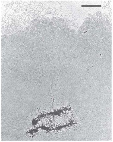
Full relaxation of the supercoils requires 1 nick/85 kb or so, thus identifying the average length of torsionally “closed” DNA. This region could comprise a loop or domain similar in nature to those identified in the bacterial genome. Loops can be seen directly when the majority of proteins are extracted from mitotic chromosomes.
The resulting complex consists of the DNA associated with about 8% of the original protein content. As shown in FIGURE 1, the protein-depleted chromosomes reveal an underlying structure of a metaphase scaffold that still resembles the general form of a mitotic chromosome, surrounded by a halo of DNA.

FIGURE 1 Histone-depleted chromosomes consist of a protein scaffold to which loops of DNA are anchored.
Reprinted from: Paulson, J. R., and Laemmli, U. K. 1977. “The structure of histonedepleted metaphase chromosomes.” Cell 12:817–828., with permission from Elsevier (http://www.sciencedirect.com/science/article/pii/009286747790280X). Photo courtesy of Ulrich K. Laemmli, University of Geneva, Switzerland.
The metaphase scaffold consists of a dense network of fibers. Threads of DNA emanate from the scaffold, apparently as loops of average length 10 to 30 μm (30 to 90 kb). The DNA can be
digested without affecting the integrity of the primarily proteinaceous scaffold. In interphase nuclei, this underlying proteinaceous structure is less well defined, but a more broadly dispersed arrangement in the nucleoplasm has been referred to as the nuclear matrix rather than the scaffold.
 الاكثر قراءة في مواضيع عامة في الاحياء الجزيئي
 اخر الاخبار
اخبار العتبة العباسية المقدسة

الآخبار الصحية


 قسم الشؤون الفكرية يصدر كتاباً يوثق تاريخ السدانة في العتبة العباسية المقدسة
 "المهمة".. إصدار قصصي يوثّق القصص الفائزة في مسابقة فتوى الدفاع المقدسة للقصة القصيرة
 (نوافذ).. إصدار أدبي يوثق القصص الفائزة في مسابقة الإمام العسكري (عليه السلام)


