آخر المواضيع المضافة

 النبات
الحيوان
الأحياء المجهرية
علم الأمراض
التقانة الإحيائية
التقنية الحيوية المكروبية
التقنية الحياتية النانوية
علم الأجنة
الأحياء الجزيئي
علم وظائف الأعضاء
الغدد
المضادات الحيوية
النبات
الحيوان
الأحياء المجهرية
علم الأمراض
التقانة الإحيائية
التقنية الحيوية المكروبية
التقنية الحياتية النانوية
علم الأجنة
الأحياء الجزيئي
علم وظائف الأعضاء
الغدد
المضادات الحيوية| Diagnostic techniques used in pathology |


|
|
Read More
Date: 28-2-2016
Date: 28-2-2016
Date: 24-2-2016
|
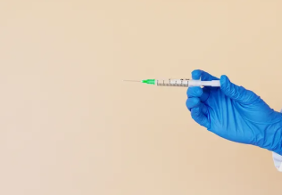
Diagnostic techniques used in pathology
The pathologist uses the following techniques to the diagnose diseases:
a. Histopathology
b. Cytopathology
c. Hematopathology
d. Immunohistochemistry
e. Microbiological examination
f. Biochemical examination
g. Cytogenetics
h. Molecular techniques
i. Autopsy
A. Histopathological techniques
Histopathological examination studies tissues under the microscope. During this study, the pathologist looks for abnormal structures in the tissue.
Tissues for histopathological examination are obtained by biopsy. Biopsy is a tissue sample from a living person to identify the disease. Biopsy can be either incisional or excisional.
Once the tissue is removed from the patient, it has to be immediately fixed by putting it into adequate amount of 10% Formaldehyde (10% formalin) before sending it to the pathologist.
The purpose of fixation is:
1. to prevent autolysis and bacterial decomposition and putrefaction
2. to coagulate the tissue to prevent loss of easily diffusible substances
3. to fortify the tissue against the deleterious effects of the various stages in the preparation of sections and tissue processing.
4. to leave the tissues in a condition which facilitates differential staining with dyes and other reagents.
Once the tissue arrives at the pathology department, the pathologist will exam it macroscopically (i.e. naked-eye examination of tissues).
Then the tissue is processed to make it ready for microscopic examination. The whole purpose of the tissue processing is to prepare a very thin tissue (i.e. five to seven μm or one cell thick tissue) which can be clearly seen under the microscope. The tissue is processed by putting it into different chemicals. It is then impregnated (embedded) in paraffin, sectioned (cut) into thin slices, & is finally stained. The stains can be Hematoxylin/Eosin stain or special stains such as PAS, Immunohistochemistry, etc...
The Hematoxylin/Eosin stain is usually abbreviated as H&E stain. The H&E stain is routinely used. It gives the nucleus a blue color & the cytoplasm & the extracellular matrix a pinkish color. Then the pathologist will look for abnormal structures in the tissue. And based on this abnormal morphology he/she will make the diagnosis. Histopathology is usually the gold standard for pathologic diagnosis.
B. Cytopathologic techniques
Cytopathology is the study of cells from various body sites to determine the cause or nature of disease.
Applications of cytopathology:
The main applications of cytology include the following:
1. Screening for the early detection of asymptomatic cancer
For example, the examination of scrapings from cervix for early detection and prevention of cervical cancer.
2. Diagnosis of symptomatic cancer
Cytopathology may be used alone or in conjunction with other modalities to diagnose tumors revealed by physical or radiological examinations.
It can be used in the diagnosis of cysts, inflammatory conditions and infections of various organs.
3. Surveillance of patients treated for cancer
For some types of cancers, cytology is the most feasible method of surveillance to detect recurrence. The best example is periodic urine cytology to monitor the recurrence of cancer of the urinary tract.
Advantages of cytologic examination
Compared to histopathological technique it is cheap, takes less time and needs no anesthesia to take specimens. Therefore, it is appropriate for developing countries with limited resources like Ethiopia. In addition, it is complementary to histopathological examination.
Cytopathologic methods
There are different cytopathologic methods including:
1. Fine-needle aspiration cytology (FNAC)
In FNAC, cells are obtained by aspirating the diseased organ using a very thin needle under negative pressure. Virtually any organ or tissue can be sampled by fine-needle aspiration. The aspirated cells are then stained & are studied under the microscope. Superficial organs (e.g. thyroid, breast, lymph nodes, skin and soft tissues) can be easily aspirated. Deep organs, such as the lung, mediastinum, liver, pancreas, kidney, adrenal gland, and retroperitoneum are aspirated with guidance by fluoroscopy, ultrasound or CT scan. FNAC is cheap, fast, & accurate in diagnosing many diseases.
2. Exfoliative cytology
Refers to the examination of cells that are shed spontaneously into body fluids or secretions. Examples include sputum, cerebrospinal fluid, urine, effusions in body cavities (pleura, pericardium, peritoneum), nipple discharge and vaginal discharge.
3. Abrasive cytology
Refers to methods by which cells are dislodged by various tools from body surfaces (skin, mucous membranes, and serous membranes). E.g. preparation of cervical smears with a spatula or a small brush to detect cancer of the uterine cervix at early stages. Such cervical smears, also called Pap smears, can significantly reduce the mortality from cervical cancer. Cervical cancer is the most common cancer in Ethiopian women.
C. Hematological examination
This is a method by which abnormalities of the cells of the blood and their precursors in the bone marrow are investigated to diagnose the different kinds of anemia & leukemia.
D. Immunohistochemistry
This is a method is used to detect a specific antigen in the tissue in order to identify the type of disease.
E. Microbiological examination
This is a method by which body fluids, excised tissue, etc. are examined by microscopical, cultural and serological techniques to identify micro-organisms responsible for many diseases.
F. Biochemical examination
This is a method by which the metabolic disturbances of disease are investigated by assay of various normal and abnormal compounds in the blood, urine, etc.
G. Clinical genetics (cytogenetics),
This is a method in which inherited chromosomal abnormalities in the germ cells or acquired chromosomal abnormalities in somatic cells are investigated using the techniques of molecular biology.
H. Molecular techniques
Different molecular techniques such as fluorescent in situ hybridization, Southern blot, etc... can be used to detect genetic diseases.
I. Autopsy
Autopsy is examination of the dead body to identify the cause of death. This can be for forensic or clinical purposes.
The relative importance of each of the above disciplines to our understanding of disease varies for different types of diseases. For example, in diabetes mellitus, biochemical investigation provides the best means of diagnosis and is of greatest value in the control of the disease. Whereas in the diagnosis of tumors, FNAC & histopathology contribute much. However, for most diseases, diagnosis is based on a combination of pathological investigations.
References
Bezabeh ,M. ; Tesfaye,A.; Ergicho, B.; Erke, M.; Mengistu, S. and Bedane,A.; Desta, A.(2004). General Pathology. Jimma University, Gondar University Haramaya University, Dedub University.


|
|
|
|
صنع الذكريات والتفكير يدمر الدماغ.. دراسة تشرح السبب
|
|
|
|
|
|
|
الصين.. عودة كاسحتي الجليد إلى شنغهاي بعد انتهاء بعثة استكشافية إلى القطب الجنوبي
|
|
|
|
|
|
جامعة الكفيل تكرم الفائزين بأبحاث طلبة كلية الصيدلة وطب الأسنان
|
|
|
|
مشروع التكليف الشرعي بنسخته السادسة الورود الفاطمية... أضخم حفل لفتيات كربلاء
|
|
|
|
ضمن جناح جمعيّة العميد العلميّة والفكريّة المجمع العلمي يعرض إصداراته في معرض تونس الدولي للكتاب
|
|
|
|
جامعة الكفيل تعقد مؤتمرها الطلابي العلمي الرابع
|